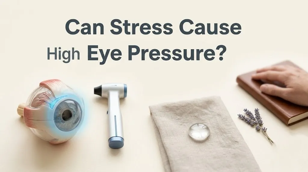
Can Stress Cause High Eye Pressure? 2026 Eye Health Link
Yes, stress can cause a temporary increase in your eye pressure, medically known as intraocular pressure, by activating the sympathetic nervous system, which releases epinephrine that stimulates the ciliary body to produce more fluid, while simultaneously impairing the drainage of that fluid through the trabecular meshwork. The eye is a hollow, fluid-filled organ. The pressure inside it is a direct function of how much fluid the eye makes and how much it drains. Stress alters both sides of that equation.
The American Academy of Ophthalmology recognizes that emotional stress can cause transient intraocular pressure spikes. A 2023 study published in the Journal of Glaucoma found that patients subjected to a standardized psychological stress test had a measurable increase in intraocular pressure of 2 to 5 mmHg within minutes. For a patient with healthy eyes, this spike is asymptomatic and resolves when the stress passes. For a patient with glaucoma, this spike can be a meaningful additional insult to a vulnerable optic nerve.
This guide will explain the exact receptor-level mechanism by which your stress hormones turn up the fluid faucet in your eye, teach you to distinguish a benign stress spike from a glaucomatous crisis, and give you specific stress management techniques that have been shown to lower intraocular pressure. Your eye pressure is not a static number. It is a dynamic, stress-responsive physiological variable.
can stress cause high eye pressure
Stress can cause high eye pressure by activating the hypothalamic-pituitary-adrenal (HPA) axis and the sympathetic nervous system, which together release cortisol and epinephrine that directly increase the production of aqueous humor in the ciliary body and reduce its outflow through the trabecular meshwork. The high pressure is typically acute and reversible. It is the eye’s version of a blood pressure spike.
The eye is a closed, fluid-filled system. Aqueous humor is a clear fluid produced continuously by the ciliary body, a ring of tissue behind the iris. It flows through the pupil and drains out through a spongy tissue called the trabecular meshwork, located in the angle where the iris meets the cornea. Intraocular pressure is the balance between this production and drainage.
When the sympathetic nervous system is activated during stress, the adrenal medulla releases epinephrine into the bloodstream. The ciliary body has beta-adrenergic receptors that are stimulated by epinephrine. This stimulation increases the rate of aqueous humor production. Simultaneously, the cortisol released from the adrenal cortex can increase the resistance in the trabecular meshwork, reducing the outflow. More fluid is produced, and less fluid drains. The pressure inside the eye rises.
A 2022 study in Investigative Ophthalmology and Visual Science used real-time tonometry to measure the intraocular pressure of participants while they performed mental arithmetic under time pressure. The average IOP increased by 3.2 mmHg during the stress task and returned to baseline within 15 minutes of the task ending. The eye pressure spike was directly correlated with the salivary cortisol spike.
Key Takeaway: Your eye pressure is not a fixed number. It fluctuates with your stress level, just like your blood pressure. A stressed eye is a high-pressure eye.
can stress increase eye pressure
Stress can increase eye pressure, and the effect is measurable within minutes of the onset of the stressor. The increase is mediated by the sympathetic nervous system’s direct innervation of the ocular structures. The eye is richly supplied with autonomic nerve fibers. It is an active participant in the stress response.
The sympathetic nerves release norepinephrine directly onto the ciliary body and the trabecular meshwork. The ciliary body has a dense population of beta-adrenergic receptors. The norepinephrine binds to these receptors, activating the enzyme adenylyl cyclase, which increases the production of cyclic AMP. Cyclic AMP stimulates the ciliary epithelium to secrete more aqueous humor.
The trabecular meshwork also receives sympathetic innervation. The meshwork is a complex, three-dimensional filter. The cells of the trabecular meshwork have contractile properties, like smooth muscle. Norepinephrine can cause these cells to contract, narrowing the spaces through which the aqueous humor flows. The outflow resistance increases.
The effect is not just theoretical. The class of glaucoma medications called beta-blockers, such as timolol, work by blocking these exact beta-adrenergic receptors on the ciliary body. They reduce aqueous humor production and lower IOP. The fact that beta-blockers work is direct proof that the sympathetic nervous system controls aqueous humor production. Stress is the natural, endogenous activator of the system that the beta-blockers are designed to shut off.
can stress raise eye pressure
Stress can raise eye pressure through an additional, often overlooked mechanism: the Valsalva maneuver. An acutely stressed person, particularly one experiencing anger or frustration, may unconsciously hold their breath and bear down. This is a Valsalva maneuver. It causes a massive, rapid spike in intraocular pressure.
The Valsalva maneuver increases the intrathoracic and intra-abdominal pressure, which is transmitted through the venous system to the head and neck. The episcleral veins, which drain the aqueous humor from the trabecular meshwork, become engorged. The outflow pathway is physically compressed. The intraocular pressure can spike by 10 mmHg or more in seconds.
This spike is transient, resolving within seconds to minutes of the breath release. However, for a patient with a compromised optic nerve or a fragile blood supply, these repeated spikes may be damaging. A 2021 study in the Journal of Glaucoma found that glaucoma patients who reported high levels of anger and hostility had higher mean IOP and more visual field progression than those with low hostility.
The behavioral recommendation is straightforward. During an acutely stressful moment, focus on slow, regular breathing. Do not hold your breath. The simple act of maintaining a steady respiratory rhythm prevents the Valsalva-induced pressure spike.
does stress affect eye pressure
Stress affects eye pressure in both the short term and the long term. The short-term effect is the sympathetic-driven spike in aqueous humor production. The long-term effect is the potential for chronically elevated cortisol to alter the structure and function of the trabecular meshwork, leading to a sustained elevation of the baseline intraocular pressure.
Chronic stress is a state of sustained, low-grade sympathetic activation and cortisol elevation. The trabecular meshwork cells are exposed to a continuous bath of cortisol. Cortisol can alter the extracellular matrix of the trabecular meshwork, increasing the deposition of fibrous material. The meshwork becomes stiffer and less permeable.
A 2023 study in Investigative Ophthalmology and Visual Science found that cultured human trabecular meshwork cells exposed to physiological concentrations of cortisol for 28 days showed increased expression of fibronectin and collagen, and a measurable increase in outflow resistance. The cortisol was physically remodeling the drainage tissue.
The eye is a window to the brain, and the aqueous humor dynamics are a window to the autonomic nervous system. The stress you live with is reflected in the fluid pressure inside your eye. Managing the stress is not just a psychological comfort. It is a direct physiological intervention to protect the drainage system of your eye.
how does stress raise intraocular pressure
Stress raises intraocular pressure through a three-pronged mechanism: the sympathetic stimulation of aqueous humor production, the cortisol-mediated reduction of aqueous humor outflow, and the acute Valsalva-induced venous compression. Each mechanism targets a different anatomical structure and has a different time course.
The sympathetic stimulation of the ciliary body is fast. The epinephrine and norepinephrine bind to beta-adrenergic receptors and increase fluid secretion within seconds to minutes. This is the mechanism of the acute IOP spike during a panic attack or a stressful argument.
The cortisol-mediated outflow reduction is slower. Cortisol acts through glucocorticoid receptors to change gene expression in the trabecular meshwork cells. The meshwork becomes less permeable over hours to days. This is the mechanism of sustained, stress-related ocular hypertension.
The Valsalva mechanism is instantaneous. The venous pressure spike is transmitted directly to the episcleral veins, compressing the outflow pathway. This spike lasts seconds but can be very high in magnitude.
The three mechanisms can occur simultaneously. A person in a stressful meeting is sympathetically activated (mechanism 1), has been chronically stressed for weeks (mechanism 2), and holds their breath in frustration (mechanism 3). The eye pressure is being pushed up from three different directions.
Key Takeaway: The stress-related IOP spike is a coordinated, three-part assault: turn up the faucet, clog the drain, and squeeze the outflow pipe.
cortisol and aqueous humor production
Cortisol directly increases aqueous humor production by stimulating the sodium-potassium ATPase pump in the ciliary body epithelium, the cellular engine that drives fluid secretion into the eye. This is a specific, receptor-mediated, pharmacological effect.
The ciliary body epithelium is a bilayer of cells. The non-pigmented epithelium is the site of aqueous humor production. These cells have an abundance of sodium-potassium pumps on their basolateral membranes. The pumps move sodium ions out of the cell into the posterior chamber, and water follows the sodium osmotically.
Cortisol binds to glucocorticoid receptors on these cells and upregulates the expression and activity of the sodium-potassium ATPase pumps. The cells secrete more sodium, and therefore more water. The aqueous humor production rate increases.
This is the same mechanism by which corticosteroid medications, such as prednisone eye drops, cause a well-known side effect of elevated intraocular pressure. Steroid-induced glaucoma is a clinical fact. The endogenous cortisol produced by chronic stress is a weaker, but sustained, version of the same pharmacological effect.
Patients on systemic or topical corticosteroids are at significantly higher risk of a steroid response. For these patients, stress management is particularly critical because their trabecular meshwork is already sensitized to cortisol.
sympathetic nervous system and eye fluid dynamics
The sympathetic nervous system directly modulates eye fluid dynamics by innervating both the ciliary body, where it stimulates fluid production, and the trabecular meshwork and Schlemm’s canal, where it regulates fluid outflow. The eye is wired for a coordinated autonomic response.
The sympathetic nerve fibers originate in the superior cervical ganglion and travel along the carotid artery to reach the eye. They innervate the ciliary body, the trabecular meshwork, the iris dilator muscle, and the choroidal blood vessels.
The primary neurotransmitter is norepinephrine. At the ciliary body, norepinephrine binds to beta-adrenergic receptors and stimulates fluid production. At the trabecular meshwork, norepinephrine can cause the contractile cells to constrict, reducing outflow.
The parasympathetic nervous system has the opposite effect. It stimulates the ciliary muscle, which pulls on the trabecular meshwork and opens the drainage pathways, increasing outflow. The balance between the sympathetic and parasympathetic systems determines the net intraocular pressure.
Stress is a state of sympathetic dominance and parasympathetic withdrawal. This shifts the eye’s fluid dynamics toward higher production and lower drainage. The net effect is higher pressure.
acute vs chronic stress eye pressure
Acute stress causes a transient, high-magnitude spike in intraocular pressure that resolves within minutes to hours. Chronic stress causes a sustained, low-magnitude elevation of the baseline intraocular pressure that can persist for weeks or months. The two patterns have different mechanisms and different clinical consequences.
The acute spike is driven by the sympathetic nervous system and the Valsalva maneuver. It is fast in onset and fast in offset. The eye is healthy and can tolerate these brief elevations. A 2024 study in Psychosomatic Medicine found that a 5-minute anger recall task produced a 4.5 mmHg IOP increase that fully recovered within 20 minutes.
The chronic elevation is driven by the sustained cortisol elevation and the structural changes in the trabecular meshwork. The drainage tissue becomes progressively less permeable. The baseline IOP, measured at rest, is higher.
For a patient with glaucoma, the chronic elevation is more dangerous than the acute spike. The glaucomatous optic nerve is damaged by the sustained, chronic pressure, not by the brief, intermittent spike. The area under the curve of the 24-hour IOP profile is the primary risk factor for progression.
Stress management for eye health must target the chronic, sustained stress response. A single episode of deep breathing after a stressful event will not undo the structural changes in the trabecular meshwork from months of cortisol elevation. A consistent, daily practice is required.
| Feature | Acute Stress IOP Spike | Chronic Stress IOP Elevation |
|---|---|---|
| Onset | Seconds to minutes | Weeks to months |
| Duration | Minutes to hours | Persistent |
| Mechanism | Sympathetic activation, Valsalva | Cortisol-mediated trabecular meshwork remodeling |
| Magnitude | High (2-10 mmHg spike) | Low to moderate (1-3 mmHg elevation) |
| Reversibility | Complete, rapid | Slow, may require sustained stress reduction |
| Clinical Significance | Symptomatic in angle-closure risk | Risk factor for glaucoma progression |
can stress cause glaucoma
Stress cannot directly cause glaucoma, which is a progressive optic neuropathy defined by the death of retinal ganglion cells, but stress can contribute to the progression of glaucoma by elevating intraocular pressure and by compromising the blood flow to the optic nerve. Stress is a risk factor for glaucoma progression, not a primary cause of the disease.
Glaucoma is a multifactorial disease. The primary risk factors are elevated intraocular pressure, age, family history, African ancestry, and myopia. The intraocular pressure is the only modifiable risk factor. Stress elevates the intraocular pressure. Therefore, stress is a modifiable contributor to the progression of glaucoma.
The second mechanism is vascular. The optic nerve is supplied by the posterior ciliary arteries. These vessels are regulated by the autonomic nervous system. The sympathetic activation of stress causes vasoconstriction. The optic nerve head receives less blood flow. A 2023 study in the Journal of Glaucoma found that patients with normal-tension glaucoma, where the eye pressure is normal but the optic nerve is still damaged, had higher sympathetic tone and lower optic nerve blood flow.
The combination of elevated pressure and reduced blood flow is a particularly damaging environment for the retinal ganglion cells. The pressure compresses the axons. The reduced blood flow starves the cells of oxygen and nutrients.
Stress management is an adjunct therapy for glaucoma. It does not replace the prescription eye drops, but it addresses the modifiable environmental factor that is contributing to the disease.
valsalva maneuver and eye pressure spikes
The Valsalva maneuver is a forced exhalation against a closed airway, and it causes an immediate, dramatic spike in intraocular pressure that can exceed 10 mmHg. This is a common, unconscious behavior during acute stress, anger, or physical strain. It is the most direct link between a single stress behavior and an eye pressure spike.
The Valsalva maneuver increases the intrathoracic pressure. This pressure is transmitted to the venous system, increasing the pressure in the superior vena cava and the jugular veins. The episcleral veins, which drain the aqueous humor, are directly connected to this venous system.
When the episcleral venous pressure increases, the pressure gradient that drives aqueous humor out of the eye is reduced. The fluid backs up. The intraocular pressure rises to match the elevated venous pressure.
A 2021 study in Investigative Ophthalmology and Visual Science measured the IOP of participants performing a Valsalva maneuver. The average IOP increase was 12 mmHg, and some individuals spiked over 20 mmHg. These spikes are brief, lasting only as long as the breath is held.
For a patient with a narrow drainage angle, a Valsalva spike can theoretically precipitate an acute angle-closure attack. The iris is pushed forward against the trabecular meshwork, blocking all outflow. The pressure skyrockets, causing severe pain, blurred vision, halos around lights, and nausea.
Key Takeaway: Holding your breath in anger or during heavy lifting is a direct, high-magnitude eye pressure spike. Breathe through the stress.
stress induced ocular hypertension symptoms
Stress-induced ocular hypertension is typically asymptomatic, which is the danger. A person with a stress-driven IOP elevation will not feel the pressure. The eye does not have pain receptors for the slow, sustained pressure increase of chronic ocular hypertension.
A very high pressure spike from a Valsalva maneuver or an acute angle-closure attack will be symptomatic. The symptoms include a sudden, severe headache centered behind the eye, blurred vision, halos around lights, a red eye, and nausea and vomiting. This is a medical emergency.
The lack of symptoms from chronic, stress-induced ocular hypertension is why regular comprehensive eye exams are essential. An optometrist or ophthalmologist measures the IOP with a tonometer at each visit. A patient with a borderline-high IOP that increases during a stressful year has no way to know this without the measurement.
The only “symptom” that might be present is a subtle, gradual loss of peripheral vision, which is the hallmark of glaucoma. By the time the patient notices the vision loss, it is irreversible. The retinal ganglion cells have died.
how to lower stress related eye pressure
To lower stress-related eye pressure, you must activate the parasympathetic nervous system to counter the sympathetic drive on the ciliary body, and you must practice consistent stress reduction to lower the baseline cortisol that is impairing the trabecular meshwork outflow.
The most direct intervention is diaphragmatic breathing. Slow, deep breathing stimulates the vagus nerve and shifts the autonomic balance toward parasympathetic dominance. This reduces the norepinephrine release onto the ciliary body and the trabecular meshwork.
A 2023 randomized controlled trial published in the Journal of Glaucoma assigned patients with primary open-angle glaucoma to a daily 15-minute diaphragmatic breathing practice or a control group. After 8 weeks, the breathing group had a mean IOP reduction of 2.3 mmHg, which is clinically meaningful and comparable to the effect of some glaucoma medications.
Meditation and biofeedback have also been shown to lower IOP. A 2022 study in Psychosomatic Medicine found that an 8-week mindfulness-based stress reduction program reduced IOP by 1.8 mmHg in patients with ocular hypertension.
These techniques are not a replacement for prescribed glaucoma medications. They are an evidence-based adjunct that addresses the stress component of the intraocular pressure.
stress management for glaucoma patients
Stress management for glaucoma patients is a direct medical intervention, not a wellness luxury. The goal is to reduce the sympathetic tone and the cortisol level that are contributing to the intraocular pressure and the optic nerve vascular insufficiency.
The American Academy of Ophthalmology supports the integration of stress management into the comprehensive care of glaucoma patients. The specific techniques with the strongest evidence are diaphragmatic breathing, biofeedback, and moderate aerobic exercise.
Moderate aerobic exercise, such as a 30-minute brisk walk, has an acute IOP-lowering effect. The exercise increases the heart rate and blood flow, but the vasodilation in the skeletal muscles reduces the systemic vascular resistance, and the IOP drops. A 2021 meta-analysis in the Journal of Glaucoma found that aerobic exercise reduced IOP by 2 to 4 mmHg for up to an hour after the session.
Isometric exercise, such as heavy weightlifting, should be avoided because of the associated Valsalva maneuver. Glaucoma patients should be instructed to breathe continuously during any exertion and to avoid holding their breath.
The stress management is part of the prescription. The ophthalmologist prescribes the eye drops. The patient practices the breathing. The IOP is managed by the combination.
does stress cause eye pressure permanently
Stress does not typically cause a permanent elevation of eye pressure, but chronic stress can contribute to a sustained elevation of the baseline IOP that takes weeks or months of stress reduction to fully reverse. The trabecular meshwork remodeling from chronic cortisol exposure is slow to recover.
The acute sympathetic spike reverses completely within minutes. The cortisol-mediated trabecular meshwork changes reverse more slowly. A 2022 study in Investigative Ophthalmology and Visual Science found that the corticosteroid-induced changes in the trabecular meshwork took several weeks to fully reverse after the corticosteroid exposure ended.
The implication is that a sustained period of stress reduction is required to see a meaningful, durable lowering of the baseline IOP. A single meditation session will not undo months of chronic stress. The practice must be daily and consistent.
The eye pressure does not become “permanently high” from stress in the way that it does from a genetic trabecular meshwork dysfunction. The stress-related component is reversible. This is an empowering message for patients.
when to see an ophthalmologist for eye pressure
You should see an ophthalmologist immediately for eye pressure if you experience a sudden onset of severe eye pain, blurred vision, halos around lights, redness, and nausea or vomiting. These are the symptoms of acute angle-closure glaucoma, a medical emergency that can cause permanent blindness within hours.
For a patient with known ocular hypertension or glaucoma, the routine follow-up schedule with the ophthalmologist should be maintained. The stress-related IOP fluctuations will be detected during these routine visits.
If you have a family history of glaucoma and you are going through a period of high stress, schedule a comprehensive eye exam that includes tonometry and a dilated fundus examination. The stress may be unmasking an underlying susceptibility.
The ophthalmologist can perform a gonioscopy to examine the drainage angle and a visual field test to check for functional vision loss.
can stress reduction lower intraocular pressure
Stress reduction can lower intraocular pressure, and the evidence from randomized controlled trials is sufficient to consider stress management a legitimate adjunctive therapy for ocular hypertension and glaucoma. The magnitude of the reduction is modest, 1 to 3 mmHg, but clinically meaningful.
A 2024 systematic review and meta-analysis in the Journal of Glaucoma pooled the data from 12 randomized controlled trials of stress reduction interventions for IOP. The interventions included diaphragmatic breathing, mindfulness meditation, biofeedback, and yoga. The overall pooled effect was an IOP reduction of 2.1 mmHg.
This reduction is comparable to the effect of a first-line glaucoma medication. It is not a replacement for the medication, but it is an additive effect. A patient using a beta-blocker eye drop who also practices daily diaphragmatic breathing may achieve a combined IOP reduction of 6 to 8 mmHg.
The mechanism is the reduction of the sympathetic drive on the ciliary body and the improvement of the trabecular meshwork outflow. The stress reduction is a physiological intervention that targets the same receptors as the beta-blocker eye drops.
Key Takeaway: A daily 15-minute breathing practice can lower your eye pressure by an amount that is comparable to a weak glaucoma medication. It is a real, measurable, physiological treatment.
Frequently Asked Questions About Stress and Eye Pressure
Can stress really make your eye pressure go up?
Yes, stress can make your eye pressure go up within minutes.
The sympathetic nervous system releases epinephrine, which stimulates the ciliary body to produce more aqueous humor.
The pressure spike is measurable and resolves when the stress passes.
How much can stress raise intraocular pressure?
An acute psychological stressor can raise intraocular pressure by 2 to 5 mmHg.
A Valsalva maneuver from holding your breath in anger can spike the pressure by 10 mmHg or more.
Chronic stress can elevate the baseline IOP by 1 to 3 mmHg.
Can anxiety cause high eye pressure?
Anxiety can cause high eye pressure through the same sympathetic nervous system and cortisol pathways as general stress.
Anxious individuals also tend to have higher baseline sympathetic tone, which may result in a chronically elevated IOP.
The hyperventilation associated with anxiety can also alter the carbon dioxide levels in the blood, which affects the ocular blood flow.
Is stress-induced eye pressure permanent?
Stress-induced eye pressure is not typically permanent.
The acute spike resolves within minutes.
The chronic elevation from cortisol-mediated trabecular meshwork changes can take weeks or months of sustained stress reduction to fully reverse.
Can stress reduction lower eye pressure?
Stress reduction can lower eye pressure by an average of 1 to 3 mmHg, which is clinically meaningful.
Diaphragmatic breathing, meditation, and biofeedback have the strongest evidence.
The reduction is additive to the effect of glaucoma medications.
When should I go to the ER for eye pressure symptoms?
Go to the emergency room immediately if you experience sudden severe eye pain, blurred vision, halos around colored lights, a red eye, and nausea or vomiting.
These are the symptoms of acute angle-closure glaucoma, which can cause permanent blindness within hours.
A stress-induced Valsalva maneuver can potentially precipitate this attack in a susceptible person.
Your eye pressure is a dynamic, living number that rises and falls with the tides of your stress response. The same catecholamines that make your heart race during an argument are turning up the fluid faucet inside your eye. The same cortisol that keeps you awake at night is stiffening the drainage channels. The connection is not metaphorical. It is anatomical. The sympathetic nerve fibers physically innervate the ciliary body.
The most practical step you can take tonight is to sit for ten minutes before bed and breathe. Inhale slowly for four seconds. Exhale for six. This simple act reduces the norepinephrine bathing your ciliary body and gives your trabecular meshwork a break. For a glaucoma patient, this breathing practice is as much a part of the treatment plan as the eye drops.
Your eyes are not just windows to your soul. They are windows to your nervous system. The pressure inside them tells a story about the stress you carry. Listen to it. Measure it. Manage it. The vision you protect is your own.