Will a Stress Test Show Blockage? 2026 Test Accuracy Guide
Yes, a cardiac stress test can show the physiological effect of a significant blockage by revealing areas of the heart muscle that are not receiving enough oxygenated blood during physical exertion or pharmacological stimulation. However, the test is not a direct camera image of the artery walls. It is a functional test. It detects a blockage indirectly by observing the downstream consequence of reduced blood flow, a condition called ischemia.
The American Heart Association and the American College of Cardiology recognize stress testing as a cornerstone of the diagnostic pathway for coronary artery disease. A 2023 meta-analysis published in the Journal of the American College of Cardiology confirmed that stress testing has a sensitivity of approximately 85 to 90 percent for detecting flow-limiting blockages. This means a normal stress test is highly reassuring, but it does not perfectly rule out all forms of coronary disease.
This guide will explain the exact physiological mechanism by which a stress test “sees” a blockage, compare the accuracy of the different types of stress tests, and clarify the critical distinction between a functionally significant blockage that shows up on a stress test and an anatomically visible blockage that might not. If you are anxious about an upcoming test or confused by a result, this is your roadmap.
If you are in crisis or experiencing thoughts of self-harm, contact the 988 Suicide and Crisis Lifeline by calling or texting 988 at any time. The fear of heart disease is real and distressing. This service is free, confidential, and available 24 hours a day.
will a stress test show blockage
A stress test will show a blockage if that blockage is hemodynamically significant, meaning it narrows a coronary artery by roughly 70 percent or more and limits the increase in blood flow that normally accompanies exercise or pharmacological stress. It will not show a mild, non-obstructive blockage of 30 or 40 percent because the blood flow through that artery can still increase adequately to meet the oxygen demand of the working heart muscle.
The heart is a muscle that requires a constant supply of oxygenated blood, delivered through the coronary arteries. At rest, the heart extracts nearly all the oxygen from the blood, so the only way to increase oxygen delivery during exertion is to increase the blood flow. Healthy coronary arteries can dilate and increase their flow by 3 to 5 times.
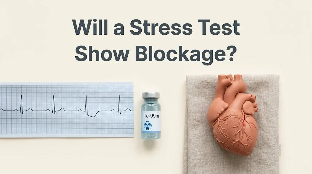
A blocked artery, stiffened and narrowed by atherosclerotic plaque, cannot dilate to the same degree. The downstream heart muscle becomes ischemic, starved for oxygen. The ischemic muscle contracts less vigorously. The EKG shows characteristic changes. The patient may feel chest pain or shortness of breath.
The stress test detects this cascade. It detects the consequence of the blockage, not the plaque itself. A 2024 review in Circulation emphasized that stress testing is a test of coronary flow reserve, the ability of the arteries to increase flow on demand. A blockage that impairs this flow reserve is the blockage the test is designed to find.
Key Takeaway: A stress test is a test of blood flow, not a picture of the artery. It finds blockages that are big enough to starve the heart muscle of oxygen during stress.
can a stress test detect blocked arteries
A stress test can detect blocked arteries that are causing a flow limitation, but it cannot directly visualize the arterial anatomy or measure the exact percentage of the stenosis. The test answers the functional question: is the blood supply to the heart muscle adequate under stress?
The detection is indirect. A nuclear stress test or a cardiac MRI perfusion scan creates a map of the blood flow to the left ventricle. A blocked artery produces a cold spot on the scan. A stress echocardiogram shows a wall motion abnormality, a segment of the heart muscle that is not squeezing as well as it should during stress.
The sensitivity of the test depends on the type of stress test and the population tested. A standard exercise EKG stress test, without imaging, has a sensitivity of about 65 to 70 percent. Adding an imaging modality, such as a nuclear perfusion scan or an echocardiogram, increases the sensitivity to approximately 85 to 90 percent.
The test is more accurate at detecting single-vessel disease than multi-vessel disease. This is a critical limitation. A patient with severe blockages in all three major coronary arteries may have a “balanced” reduction in flow to all areas of the heart. No region is relatively worse than another. The perfusion map looks normal. The stress test is falsely negative.
how does a stress test show blockages
A stress test shows blockages by creating a controlled state of increased myocardial oxygen demand, usually through treadmill exercise or a pharmacological agent like dobutamine, and then comparing the heart’s function and blood flow at rest to its function and blood flow during peak stress. The difference between the resting and stressed states reveals the presence and location of flow-limiting blockages.
The mechanism of exercise stress is the most physiological. The patient walks or runs on a treadmill, and the workload is progressively increased. The heart rate and the blood pressure rise. The myocardial oxygen demand increases. The healthy coronary arteries dilate to meet the demand. A diseased artery cannot.
The EKG monitors the electrical activity of the heart. Ischemia produces a characteristic depression of the ST segment on the EKG tracing. The depth and duration of the ST depression correlate with the severity of the ischemia.
The imaging component adds spatial information. A nuclear tracer, such as technetium-99m sestamibi, is injected at peak stress. The tracer is taken up by the heart muscle cells in proportion to the blood flow they receive. A gamma camera then creates a three-dimensional map of the perfusion. A stress-induced perfusion defect is a dark spot on the scan where the blocked artery has limited the tracer uptake.
will a stress test show a blockage
A stress test will show a blockage that is causing a significant flow reduction, but the specific phrasing “show a blockage” requires clarification. The test shows the physiological effect of a blockage. It does not show the blockage itself in the way a coronary CT angiogram or an invasive angiogram shows the plaque.
The stress test result is reported as normal, equivocal, or abnormal. An abnormal result indicates a high probability of a flow-limiting blockage. The report will often specify the likely location of the blockage based on which wall of the heart is affected. A defect in the anterior wall suggests a blockage in the left anterior descending artery.
The magnitude of the abnormality is also reported. A small, mild defect is less concerning than a large, severe defect. A transient ischemic dilation, where the left ventricle appears larger on the stress images than the rest images, is a high-risk marker for severe multi-vessel disease.
The result is not a percentage stenosis. It is a functional assessment. The next step after an abnormal stress test is often a coronary angiogram, which directly visualizes the artery and measures the anatomical narrowing.
how accurate is a nuclear stress test
A nuclear stress test, also called a myocardial perfusion imaging test, has a sensitivity of approximately 85 to 90 percent and a specificity of approximately 70 to 80 percent for detecting flow-limiting coronary artery disease. The accuracy is higher than a standard exercise EKG test and comparable to a stress echocardiogram.
The sensitivity is the ability of the test to correctly identify a patient who has the disease. A sensitivity of 88 percent means that out of 100 patients with a flow-limiting blockage, 88 will have an abnormal nuclear stress test. Twelve will have a false-negative test.
The specificity is the ability of the test to correctly identify a patient who does not have the disease. A specificity of 75 percent means that out of 100 patients without a blockage, 75 will have a normal test. Twenty-five will have a false-positive test.
A 2023 study in the Journal of the American College of Cardiology compared the diagnostic accuracy of nuclear stress testing to the gold standard of invasive fractional flow reserve, or FFR. The nuclear stress test had an area under the receiver operating characteristic curve of 0.89, indicating excellent diagnostic performance.
The accuracy can be reduced by artifacts. Breast tissue and the diaphragm can absorb the gamma rays and create an apparent defect on the scan, particularly in women and obese patients.
Key Takeaway: A nuclear stress test is about 90 percent sensitive. It misses about 10 percent of significant blockages. It is not a perfect test, but it is a very good one.
does a stress test show blocked arteries
A stress test shows the functional consequence of a blocked artery, not the artery itself. The report will state whether there is evidence of ischemia, not whether there is a 50 percent or 80 percent plaque. The distinction is important.
A patient can have a 50 percent plaque on an angiogram and a normal stress test because the artery can still dilate enough to maintain adequate flow during stress. This plaque is anatomically present but functionally silent. Medical therapy, not a stent, is the appropriate treatment.
Conversely, a patient can have a normal angiogram and an abnormal stress test. This is microvascular disease, where the large arteries are open but the tiny resistance vessels are dysfunctional and cannot dilate. The stress test detects the microvascular ischemia.
The stress test is a functional test. The angiogram is an anatomical test. They provide complementary, not identical, information.
types of cardiac stress tests for blockage
There are four primary types of cardiac stress tests used to detect blockages: the exercise EKG stress test, the stress echocardiogram, the nuclear stress test, and the cardiac MRI stress test. Each has a different mechanism of detecting ischemia and a different accuracy profile.
The exercise EKG stress test is the simplest. The patient walks on a treadmill, and the EKG is monitored. It detects ischemia by ST segment depression. Sensitivity is 65 to 70 percent. It is a good initial screening test for patients with a normal baseline EKG who can exercise adequately.
The stress echocardiogram uses ultrasound to visualize the heart’s wall motion at rest and immediately after peak stress. A wall motion abnormality during stress indicates ischemia. Sensitivity is 80 to 85 percent. It avoids radiation exposure.
The nuclear stress test uses a radioactive tracer to map blood flow. It provides the highest degree of quantification of the ischemia. Sensitivity is 85 to 90 percent. The radiation dose is approximately 8 to 15 mSv.
The cardiac MRI stress test is the newest and most accurate. It uses a gadolinium contrast agent to map perfusion and can also visualize the scar from a previous heart attack. Sensitivity is 90 to 95 percent. It is the most expensive and least available.
| Test Type | Mechanism | Sensitivity | Radiation | Best For |
|---|---|---|---|---|
| Exercise EKG | ST segment changes on EKG | 65-70% | None | Initial screen, normal baseline EKG |
| Stress Echo | Wall motion abnormality on ultrasound | 80-85% | None | Patients who can exercise, need no radiation |
| Nuclear Stress Test | Perfusion defect on SPECT or PET scan | 85-90% | 8-15 mSv | Quantifying ischemia extent |
| Cardiac MRI Stress | Perfusion defect on MRI | 90-95% | None | Highest accuracy, complex cases |
stress test vs angiogram for blockage detection
A stress test detects a functionally significant blockage by demonstrating ischemia, while an angiogram detects an anatomically significant blockage by directly visualizing the narrowing of the artery lumen. The stress test answers the question, “Is the blockage causing a problem?” The angiogram answers the question, “What is the exact location and severity of the blockage?”
A coronary angiogram is an invasive procedure where a catheter is threaded through an artery in the wrist or groin to the coronary arteries. Contrast dye is injected, and X-ray images are taken. The cardiologist measures the percentage of the artery that is narrowed.
The angiogram is the gold standard for anatomical diagnosis. However, it has a limitation. It cannot always determine whether an intermediate blockage, between 40 and 70 percent, is functionally significant. A fractional flow reserve, or FFR, measurement can be done during the angiogram to answer this.
The stress test and the angiogram are sequential steps in a diagnostic pathway. An abnormal stress test leads to an angiogram. A normal stress test usually means an angiogram is not necessary.
what does a stress test show about your heart
A stress test shows your heart’s exercise capacity, the presence or absence of inducible ischemia, the global function of your left ventricle, your heart rate and blood pressure response to exercise, and the presence of any exercise-induced arrhythmias. It is a comprehensive cardiovascular performance assessment.
The exercise capacity is measured in metabolic equivalents, or METs. A higher MET level indicates better cardiovascular fitness. The MET level achieved is a powerful independent predictor of survival.
The stress test also shows the location and extent of any ischemia. A small, mild defect in a single territory is low risk. A large, severe defect in multiple territories is high risk.
The test also provides information about the heart’s electrical stability. Ventricular ectopy that worsens with exercise is a marker of increased risk.
can a stress test miss a blockage
A stress test can miss a blockage in several specific scenarios. The most important is balanced ischemia, where severe three-vessel disease produces a globally reduced flow that appears normal on a relative perfusion scan.
A stress test can also miss a blockage if the patient does not achieve an adequate level of cardiac stress. The target heart rate is 85 percent of the age-predicted maximum. If the test is stopped early due to fatigue or the medication is insufficient, the sensitivity drops.
A stress test can miss a non-flow-limiting blockage. A 50 percent stenosis in the left anterior descending artery that has a normal fractional flow reserve will not cause ischemia and will not be detected.
A stress test can be falsely normal due to technical factors, such as attenuation artifact obscuring a true defect.
stress test false positive for blockage
A stress test can be falsely positive for a blockage, meaning the test is abnormal but the patient does not have significant coronary artery disease on an angiogram. False positives are more common in certain populations.
Breast tissue attenuation in women is a common cause of a false-positive nuclear stress test. The breast tissue absorbs the gamma rays and creates an apparent defect in the anterior wall of the heart. Diaphragmatic attenuation can create an inferior wall defect.
A left bundle branch block on the EKG can produce a false-positive septal perfusion defect on a nuclear stress test, even in the absence of a blockage.
A hypertensive response to exercise can produce EKG changes that mimic ischemia.
A false-positive test can cause significant anxiety and lead to an unnecessary invasive angiogram. The pre-test probability of disease should always be considered when interpreting a stress test result.
functional vs anatomical coronary blockage
A functional coronary blockage is a narrowing that is severe enough to impair the increase in blood flow during stress, causing ischemia. An anatomical coronary blockage is a visible narrowing of the artery on an angiogram, regardless of whether it impairs flow. The distinction defines the treatment.
An anatomical 50 percent stenosis is visible. It is often not flow-limiting. The stress test will be normal. Stenting this lesion does not improve outcomes. Medical therapy is the standard of care.
A functional blockage, typically a 70 percent or greater stenosis, impairs coronary flow reserve. The stress test is abnormal. Revascularization with a stent or bypass surgery, combined with medical therapy, may be indicated.
The ISCHEMIA trial, a landmark 2020 study published in the New England Journal of Medicine, demonstrated that for patients with stable coronary artery disease and moderate to severe ischemia on a stress test, an initial invasive strategy of stenting or bypass surgery did not reduce the risk of death or heart attack compared to optimal medical therapy alone. The trial reinforced the importance of functional testing and the need for nuanced decision-making.
Key Takeaway: The stress test detects the functional blockage, the one that is starving the heart muscle. The angiogram sees all the blockages, big and small. Treating the functional ones is what matters for symptoms.
stress test for microvascular disease
A stress test can detect microvascular disease, where the large epicardial coronary arteries are open but the tiny resistance vessels are dysfunctional and cannot dilate. This condition, called coronary microvascular dysfunction, is more common in women and is a cause of chest pain with normal angiograms.
The nuclear stress test or cardiac MRI may show a globally reduced coronary flow reserve, even though the perfusion images do not show a localized defect. The stress echocardiogram may show a global reduction in wall motion thickening.
The diagnosis is made by measuring the coronary flow reserve on a PET stress test or by performing an invasive coronary reactivity test during an angiogram.
Microvascular disease is a real, organic cause of ischemia. It is not “just anxiety.” The treatment involves aggressive risk factor control, beta-blockers, and sometimes medications like ranolazine.
when to get a stress test for blockage symptoms
You should get a stress test when you have symptoms suggestive of coronary artery disease, such as exertional chest pain or pressure, unexplained shortness of breath with activity, or a significant decrease in your exercise tolerance, particularly if you have risk factors like diabetes, hypertension, or a strong family history of early heart disease.
The American Heart Association guidelines recommend stress testing for patients with an intermediate pre-test probability of coronary artery disease. This is a clinical judgment based on age, sex, and symptom characteristics.
A stress test is not recommended as a routine screening test for asymptomatic individuals with low risk. The false-positive rate in a low-risk population is high, and the downstream testing can cause more harm than benefit.
A primary care physician or a cardiologist should determine the appropriateness of the test based on a detailed clinical history.
preparing for a stress test to detect blockages
To prepare for a stress test, you must fast for 4 to 6 hours before the test, avoid caffeine for 12 to 24 hours because it blocks the receptors for the pharmacological stress agents, and hold specific cardiac medications as directed by your physician.
Caffeine is a methylxanthine that blocks the adenosine receptors. If you consume caffeine before a pharmacological nuclear stress test, the medication will not produce adequate vasodilation, and the test will be non-diagnostic.
Beta-blockers slow the heart rate. They are often held for 24 to 48 hours before an exercise stress test so the patient can achieve an adequate chronotropic response.
Wear comfortable shoes and clothing for the treadmill. Do not apply lotion or powder to your chest.
what happens after an abnormal stress test
After an abnormal stress test, the next step is typically a referral to a cardiologist for further evaluation, which may include a coronary CT angiogram to directly visualize the arteries or an invasive coronary angiogram in the cardiac catheterization laboratory.
The decision depends on the severity and extent of the abnormality. A small, mild defect may be managed with medical therapy and risk factor modification. A large, severe defect warrants an invasive angiogram.
The cardiologist will review the stress test images and the clinical history and discuss the next steps. The invasive angiogram is the definitive test for visualizing the coronary anatomy and guiding the need for revascularization.
An abnormal stress test is not a reason to panic. It is a piece of data that guides the next step in a well-established diagnostic pathway.
Frequently Asked Questions About Stress Tests and Blockages
Can a stress test show if you have blocked arteries?
A stress test can show if you have blocked arteries that are causing a significant reduction in blood flow to the heart muscle during exertion.
It detects the functional consequence of the blockage, not the plaque itself.
A normal test is reassuring but does not rule out small, non-flow-limiting blockages.
How accurate is a nuclear stress test for blockages?
A nuclear stress test has a sensitivity of about 85 to 90 percent and a specificity of about 70 to 80 percent for detecting flow-limiting blockages.
It is more accurate than a standard treadmill test without imaging.
Artifacts and balanced ischemia can reduce its accuracy.
What if my stress test is normal but I still have pain?
If your stress test is normal but you still have pain, the pain may be non-cardiac, or you may have coronary microvascular dysfunction.
Microvascular disease affects the tiny resistance vessels and is more common in women.
A PET stress test or an invasive coronary reactivity test can diagnose this condition.
Can anxiety cause a stress test to be abnormal?
Anxiety can cause changes on the EKG during a stress test, particularly ST segment flattening, which can be mistaken for ischemia.
Hyperventilation from anxiety can also produce EKG changes.
The imaging component of the test helps to differentiate a true perfusion defect from an anxiety-related EKG change.
What is the best test to see blocked arteries?
The best test to directly see the anatomy of blocked arteries is a coronary CT angiogram or an invasive coronary angiogram.
A stress test is the best test to determine if a blockage is functionally significant and causing ischemia.
The two types of tests provide complementary information.
Will a stress test miss a blockage?
A stress test can miss a blockage if it is non-flow-limiting, if the patient does not achieve an adequate level of stress, or in the case of balanced ischemia.
Balanced ischemia occurs when all three coronary arteries are severely narrowed, and the perfusion is globally reduced rather than regionally.
A coronary CT angiogram can detect these blockages.
The cardiac stress test is a brilliant and widely misunderstood diagnostic tool. It does not take a photograph of your coronary arteries. It measures the physiological reserve of your heart muscle. It asks the simple, elegant question: can your heart’s blood supply increase to meet the increased demand of physical stress? A significant blockage is a “no” answer to that question.
If you are scheduled for a stress test, your job is to show up prepared, fasted, and caffeine-free, and to push yourself on the treadmill to the best of your ability. The harder you work, the more accurate the test. If you are anxious about the result, know that a normal stress test is powerfully reassuring, and an abnormal one is a clear roadmap to the next step.
Your heart is a muscle with a remarkable capacity to adapt. The stress test is a snapshot of its current functional status. Treat the results as information, not a verdict. And remember to breathe. The psychological stress you bring to the test is not the stress being measured, but it can still affect your heart rate.