Neck Stress and Vertigo: The Connection Explained 2026
Yes, neck stress can cause vertigo, and the mechanism is specific: psychological stress activates your sympathetic nervous system and HPA axis, releasing cortisol and norepinephrine that increase resting muscle tension in your cervical spine, particularly in the suboccipital muscles at the base of your skull, which distorts proprioceptive signals to your brainstem and creates a sensory mismatch you perceive as dizziness or spinning.
Research published in the Journal of Vestibular Research confirms that the cervical spine contains one of the densest concentrations of proprioceptors in the human body, with approximately 200 muscle spindles per gram of tissue in the suboccipital muscles compared to roughly 16 per gram in larger limb muscles. When stress keeps these muscles chronically tight, the distorted signals they send to your vestibular nuclei create real balance disruption, not imagined symptoms.
This article explains the exact biological pathway from a stressful thought to a dizzy spell, gives you a clear framework for distinguishing stress-related cervical vertigo from inner ear disorders like BPPV or Meniere’s disease, and provides specific techniques to address both the stress and the neck tension. You will learn which stretches actually target the muscles causing the problem, when self-management is enough, and exactly when a vestibular physical therapist or ENT specialist needs to take over.
Can Neck Stress Cause Vertigo
Neck stress causes vertigo through a chain reaction that begins with psychological stress activating your autonomic nervous system, progresses to chronic muscle hypertonicity in the cervical spine, and ends with disrupted proprioceptive signaling to the vestibular nuclei in your brainstem, creating a mismatch between what your neck sensors report and what your inner ear detects.
This is not a vague association. Your brain constantly integrates three streams of sensory information to maintain balance: visual input from your eyes, vestibular input from your inner ear, and proprioceptive input from your joints and muscles, particularly your neck. The cervical spine’s proprioceptors tell your brain where your head is positioned relative to your body. When stress keeps cervical muscles in a state of sustained contraction, those proprioceptors fire inaccurately. Your brain receives a conflict between neck signals and inner ear signals, and that conflict produces dizziness.
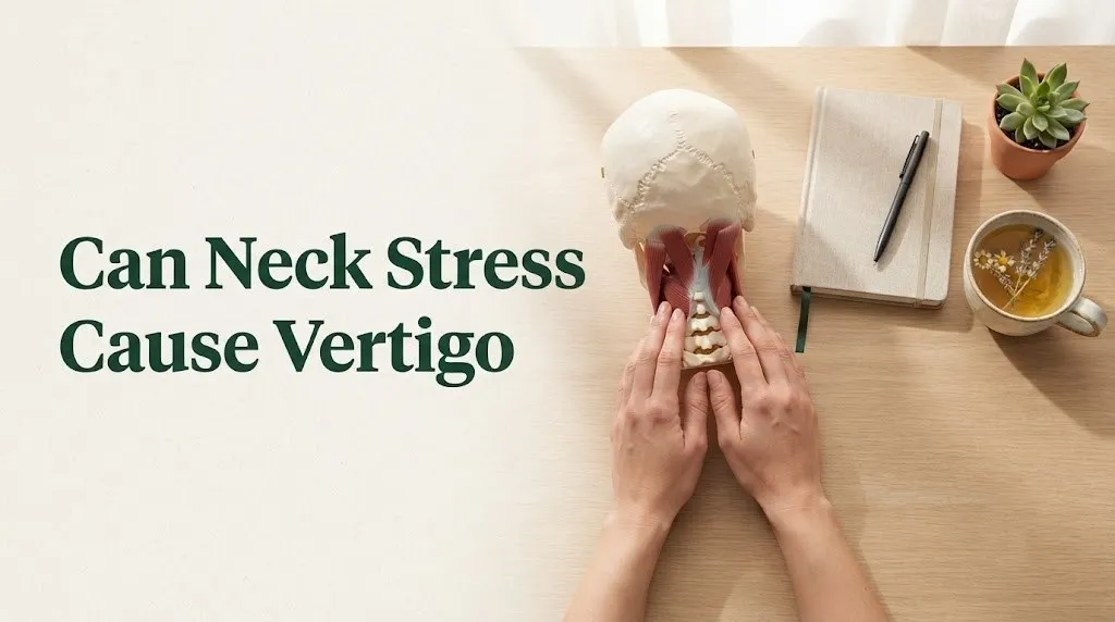
The Vestibular Disorders Association recognizes cervicogenic dizziness as a distinct clinical entity, separate from vertigo caused by inner ear disorders. The key diagnostic feature is that the dizziness is provoked or worsened by neck positions or movements, and it improves when the cervical dysfunction is treated. For the person experiencing it, this means the vertigo is real and physiologically based, even though the root cause begins with stress rather than structural damage.
Key finding: A 2022 study published in the Journal of Orthopaedic and Sports Physical Therapy found that patients with chronic neck pain and dizziness showed significantly elevated scores on the Perceived Stress Scale compared to those with neck pain alone, suggesting stress is an independent amplifier of cervical-vestibular dysfunction.
What Is Cervical Vertigo
Cervical vertigo, formally called cervicogenic dizziness, is a type of dizziness or imbalance caused by dysfunction in the cervical spine that disrupts the proprioceptive information traveling from neck muscles and joints to the brainstem’s balance centers. It is distinct from vertigo caused by inner ear problems because the source is in the neck, not the vestibular apparatus itself.
The condition was first described in the medical literature in the 1950s when researchers noticed that patients with whiplash injuries and cervical arthritis often reported dizziness that could not be explained by ear examination. Since then, the understanding has expanded to include non-traumatic causes, with stress-induced muscle tension recognized as a trigger for the same proprioceptive disruption that trauma can produce. The pathophysiology is the same: altered sensory input from cervical structures to the vestibular nuclei. The difference is the cause of that altered input: stress and muscle tension rather than structural damage from injury or degeneration.
Despite being a recognized clinical entity, cervical vertigo remains underdiagnosed. The symptoms overlap with more common causes of dizziness, and many healthcare providers default to inner ear diagnoses without evaluating the cervical spine. The American Physical Therapy Association’s vestibular rehabilitation guidelines include specific cervical screening for patients with dizziness, recommending that neck position testing be part of every dizziness evaluation, particularly when the patient also reports neck pain or stiffness.
Quick Tip:
If your dizziness changes when you turn your head, look up or down, or press on the muscles at the base of your skull, the neck is almost certainly involved. Document which movements trigger symptoms before your medical appointment.
How Stress Causes Neck Muscle Tension
Stress causes neck muscle tension through direct sympathetic nervous system activation that increases the baseline firing rate of motor neurons supplying cervical muscles, creating a state of sustained low-level contraction that persists as long as the stress response remains active. This is a hardwired physiological response, not a habit or posture problem alone.
When your brain perceives a stressor, the amygdala signals the hypothalamus to activate two parallel systems. The sympathetic-adrenal-medullary (SAM) axis releases norepinephrine from sympathetic nerve terminals throughout your body, including the motor neurons that control your trapezius, levator scapulae, and suboccipital muscles. Norepinephrine increases the excitability of these neurons, making the muscles contract more readily and relax less completely. Simultaneously, the HPA axis triggers cortisol release, which sensitizes muscle tissue to the effects of norepinephrine over hours to days.
The neck is particularly vulnerable to this stress-induced hypertonicity for anatomical reasons. The suboccipital muscles have the highest density of muscle spindles, specialized stretch receptors that detect changes in muscle length, of any muscles in the body. They are designed to make fine adjustments to head position. Under stress, their baseline tension increases disproportionately, and the dense spindle population means this tension produces an outsized effect on proprioceptive signaling. It is like turning up the gain on a microphone until it feeds back, except the feedback is distorted position sense rather than sound.
Individual variation note: People with pre-existing anxiety disorders often show higher baseline cervical muscle tension even at rest, measured by electromyography. If you have generalized anxiety disorder or panic disorder, your neck muscles may already be operating at a higher tension set point, making you more vulnerable to stress-induced cervical vertigo at lower stress thresholds.
The Connection Between Stress Hormones and Cervical Proprioceptors
The connection between stress hormones and cervical proprioceptors operates at the cellular level: cortisol and norepinephrine alter the firing characteristics of muscle spindles and Golgi tendon organs in the cervical spine, changing both the baseline discharge rate and the sensitivity of these sensory receptors to stretch and tension.
Muscle spindles are sensory organs embedded within muscle fibers that detect changes in muscle length and the rate of that change. In the suboccipital region, spindle density reaches approximately 200 spindles per gram, the highest concentration anywhere in the human body. These spindles constantly send information to the vestibular nuclei about head position relative to the trunk. When stress hormones increase the resting tension of the muscles containing these spindles, the spindles fire as if the muscle is being stretched, even when the head is in a neutral position. The brain receives signals that conflict with vestibular and visual input, and the resulting mismatch produces dizziness.
Golgi tendon organs, which detect muscle tension rather than length, are similarly affected. Elevated norepinephrine alters their firing threshold, meaning they signal “excessive tension” at lower levels of actual force. The combined effect is a proprioceptive system sending inaccurate information about both head position and neck muscle force, and the brain’s balance centers cannot reconcile this with what the inner ear reports.
Think of it this way: your brain’s balance system works like a GPS that triangulates signals from three satellites: your eyes, your inner ear, and your neck proprioceptors. Stress-induced neck tension is like one satellite suddenly broadcasting inaccurate coordinates. The GPS does not know which signal to trust, and the result is navigational confusion. Your experience of that confusion is dizziness.
Key Takeaway: Your stress hormones do not just make you feel anxious. They physically change the signals your neck muscles send to your brain’s balance centers. Treating the stress treats the signal distortion at its source.
What Does Stress-Related Neck Vertigo Feel Like
Stress-related neck vertigo typically feels like a sensation of floating, swaying, or unsteadiness rather than the intense room-spinning vertigo characteristic of inner ear disorders, and it worsens with specific neck positions, prolonged sitting, or during periods of heightened stress. People often describe it as feeling like they are walking on a boat, even when standing on solid ground.
The quality of the dizziness is distinctly different from BPPV or Meniere’s disease. With stress-induced cervical vertigo, you are more likely to experience a sense of internal motion, as if you yourself are moving, rather than the external spinning of the environment that occurs with inner ear vertigo. The sensation may be continuous but low-grade rather than episodic and severe. Many people report that the dizziness is worse after hours at a computer, when looking up at a screen or down at a phone for extended periods, or during the late afternoon and evening after a stressful workday.
Associated symptoms often include a dull ache or pressure at the base of the skull, tightness across the upper shoulders, and a feeling of heaviness or instability in the head. Some people experience mild visual disturbances, a sense that their eyes cannot track smoothly, or difficulty focusing during head movements. These visual symptoms occur because the brain uses eye movements to compensate for the distorted proprioceptive input, and the extra effort creates eye strain or blurring.
| Sensation | Cervical Vertigo (Stress) | BPPV (Inner Ear) |
|---|---|---|
| Quality of dizziness | Floating, swaying, unsteadiness | Intense room spinning (rotational) |
| Duration | Continuous or hours-long | Seconds to minutes per episode |
| Triggers | Neck position, sustained posture, stress | Specific head movements (rolling in bed) |
| Associated symptoms | Neck pain, stiffness, shoulder tension | Nausea, vomiting, nystagmus (eye jerking) |
| Effect of neck massage | Often provides temporary relief | No effect |
| Stress correlation | Strong, worsens with stress | Weak or none |
Cervical Vertigo Symptoms Checklist
Cervical vertigo presents with a recognizable pattern of symptoms that can help you identify whether your dizziness is likely neck-related before you see a healthcare provider. The presence of dizziness combined with neck pain or stiffness and symptom changes with neck position are the strongest indicators.
The Vestibular Disorders Association identifies the core feature as dizziness that is provoked or altered by neck positions or movements. This means turning your head to check a blind spot while driving, looking up at a high shelf, or tilting your head down to read may trigger or worsen the sensation. Unlike BPPV, which produces brief, intense spinning with position changes, cervical vertigo typically produces a less intense but more persistent unsteadiness that shifts with neck movement.
A second key feature is the presence of cervical spine discomfort. If you have no neck pain, tenderness, or stiffness, cervical vertigo is less likely. The neck symptoms may predate the dizziness or develop concurrently. Some people become aware of neck tension only after the dizziness starts because the dizziness draws attention to the neck. Palpating or pressing on the suboccipital muscles, the small muscles just below the skull, often reproduces or alters the dizziness, a finding known as cervicogenic dizziness provocation.
Cervical vertigo symptom checklist:
- Dizziness or unsteadiness that changes with head or neck movement
- Sensation of floating, swaying, or walking on uneven ground
- Neck pain, stiffness, or reduced range of motion accompanying the dizziness
- Symptoms worsen after prolonged sitting, especially at a computer
- Dizziness improves temporarily after neck massage, heat, or stretching
- Mild visual disturbance: difficulty focusing, eye strain with head movement
- Ear symptoms (fullness, tinnitus, hearing loss) are typically absent
- Stress level correlates with symptom severity: worse during high-stress periods
Individual variation note: People with migraine disorders may experience overlapping cervical vertigo and vestibular migraine symptoms. If you have a history of migraines and dizziness with neck pain, both mechanisms may be contributing. A neurologist or headache specialist can help differentiate and treat both components.
How to Tell If Vertigo Is From Neck Stress or Inner Ear
You can distinguish neck stress vertigo from inner ear vertigo by tracking four features: the quality of the dizziness, the duration of episodes, the triggers that provoke symptoms, and the presence or absence of ear-related symptoms like hearing loss or tinnitus. The pattern these features create points clearly toward either a cervical or vestibular origin.
Inner ear vertigo typically produces intense rotational spinning, the sensation that the room is moving around you. Episodes are usually brief, lasting seconds to minutes for BPPV, or hours for Meniere’s disease. The triggering event for BPPV is highly specific: rolling over in bed, tipping the head back, or bending forward. These movements change the position of otoconia, tiny calcium crystals in the inner ear, causing them to move within the semicircular canals and trigger a burst of false motion signals. Cervical vertigo does not follow this pattern. It changes with sustained neck positions and muscle tension levels, not with brief position changes alone.
Ear symptoms are another discriminator. Meniere’s disease includes episodic vertigo with fluctuating hearing loss, tinnitus, and a feeling of fullness in the affected ear. Vestibular migraine often includes light and sound sensitivity, visual aura, and headache. Cervical vertigo typically lacks ear-specific symptoms. If your hearing is normal, you have no tinnitus or ear fullness, and your dizziness correlates with neck discomfort and stress levels, cervical vertigo is the more likely explanation.
| Feature | Neck Stress Vertigo | BPPV | Meniere’s Disease |
|---|---|---|---|
| Dizziness quality | Swaying, floating | Intense spinning | Spinning with nausea |
| Episode duration | Hours to continuous | Under 1 minute | 20 minutes to 12+ hours |
| Key triggers | Neck position, stress, prolonged sitting | Head position changes (rolling, looking up) | Spontaneous, sometimes salt or caffeine |
| Neck pain or stiffness | Usually present | Absent | Absent |
| Hearing loss or tinnitus | Absent | Absent | Present (fluctuating) |
| Response to neck treatment | Improves | No effect | No effect |
Key Takeaway: If pressing on the muscles at the base of your skull changes your dizziness, and if neck massage or stretching provides temporary relief, the neck is the primary driver regardless of whether an inner ear issue also exists. Start with cervical treatment and stress management.
Forward Head Posture and Dizziness Connection
Forward head posture contributes to dizziness by increasing the mechanical load on the cervical spine, forcing the suboccipital muscles into constant contraction to hold the head in a forward position, and progressively distorting the proprioceptive signals these muscles send to the vestibular nuclei. Each inch your head sits forward of neutral adds approximately 10 pounds of effective weight that your neck muscles must support.
The biomechanics work against balance in two ways. First, the sustained contraction of the suboccipital muscles under increased load creates the same type of distorted proprioceptive firing that stress-induced tension produces. Even without psychological stress, forward head posture alone can generate cervical vertigo through pure mechanical overload. When you add stress-related muscle hypertonicity on top of this postural strain, the effect is compounded. Your neck muscles are simultaneously tight from stress and overloaded from posture.
Second, forward head posture alters the alignment of the cervical spine, particularly at the atlanto-occipital joint where the skull meets the first cervical vertebra. This joint has a high concentration of proprioceptors and is critical for head-on-neck position sense. Chronic malalignment changes the resting discharge pattern of these receptors, creating a persistent mismatch with vestibular input. Research published in Spine in 2023 demonstrated that participants with forward head posture showed significantly altered cervical joint position sense, the ability to accurately perceive head position, compared to those with neutral alignment.
For desk workers, the mechanism is predictable: eight hours of forward head posture at a computer creates sustained suboccipital compression. Stress from deadlines, meetings, and workload adds sympathetic-driven muscle tension. The combination distorts proprioceptive input progressively through the day. By late afternoon, the sensory mismatch reaches a threshold that produces noticeable dizziness or unsteadiness.
Suboccipital Muscle Tension and Balance Disruption
The suboccipital muscles, a group of four small muscles located at the very base of your skull, are the anatomical link between stress and vertigo because they contain the highest density of muscle spindles in the human body and their tension level directly determines the quality of proprioceptive information reaching your brainstem balance centers.
These four paired muscles, rectus capitis posterior major, rectus capitis posterior minor, obliquus capitis superior, and obliquus capitis inferior, connect the upper cervical vertebrae to the base of the skull. Their primary function is fine head position control and postural stabilization, not gross movement. They are designed for precision, which is why they have such dense sensory innervation. The rectus capitis posterior minor has an additional unique connection: it attaches directly to the dura mater, the protective covering of the brain and spinal cord, through a connective tissue bridge. Some researchers propose that tension in this muscle could directly affect dural tension and cerebrospinal fluid dynamics, though this mechanism remains under investigation.
When these muscles are chronically tight from stress, the muscle spindles fire inaccurately. Normal head movements produce exaggerated sensory signals. The vestibular nuclei receive conflicting information: the inner ear says the head is moving one way, the eyes report another, and the neck proprioceptors report a third. The brainstem resolves this conflict imperfectly, and the result is a sensation of unsteadiness or motion when no real threat to balance exists.
How to locate your suboccipital muscles:
- Place your fingers on the bony ridge at the very base of your skull, just behind your ears
- Slowly tilt your head back slightly; you will feel small muscles engage under your fingertips
- These are the suboccipitals; tenderness or tension here that reproduces dizziness confirms cervical involvement
- Gentle pressure or massage here often temporarily alters dizziness, which is diagnostically useful to note
Key Takeaway: The muscles causing your dizziness are about the size of your thumb, located exactly where your skull meets your neck. Releasing them directly is one of the fastest ways to reduce cervical vertigo symptoms, and you can learn to do it yourself.
Immediate Relief for Stress-Induced Neck Vertigo
The fastest immediate relief for stress-induced neck vertigo comes from a combination of suboccipital muscle release, gentle cervical range-of-motion exercises, and brief parasympathetic nervous system activation through controlled breathing. This three-part approach addresses the muscle tension directly, restores normal proprioceptive signaling, and reduces the sympathetic drive that maintains the tension.
Suboccipital release works by mechanically reducing the compression and hypertonicity in the muscles that are distorting proprioceptive input. When these muscles relax, the spindle firing rates normalize, and the sensory mismatch at the vestibular nuclei decreases. Many people notice a reduction in dizziness within five to ten minutes of targeted suboccipital release. The effect is temporary but confirms the cervical origin of the symptoms and provides a tool for managing acute episodes.
The parasympathetic activation component is equally important because it addresses the underlying driver. Slow, diaphragmatic breathing at five to seven breaths per minute reduces norepinephrine release from sympathetic nerve terminals. As norepinephrine levels drop, resting muscle tone decreases throughout the body, including in the cervical spine. The combination of direct mechanical release and autonomic downregulation produces faster and more complete relief than either approach alone.
For immediate relief when dizziness strikes:
- Sit in a chair with back support and place a small rolled towel behind your neck at the base of your skull
- Allow your head to rest back gently into the towel so the suboccipital muscles are supported and can release
- Begin slow nasal breathing: inhale for a count of four, exhale for a count of six
- While breathing slowly, gently turn your head from side to side about 10 to 15 degrees, staying within a pain-free range
- Continue for five to ten minutes, then reassess dizziness
- If the dizziness has decreased, the neck is confirmed as the primary driver
Safety note: If any movement significantly worsens dizziness or causes sharp pain, stop immediately. Positional vertigo that intensifies dramatically with head movement suggests BPPV rather than cervical vertigo and warrants evaluation by an otolaryngologist or vestibular physical therapist.
Neck Stretches to Reduce Cervical Dizziness
Specific neck stretches reduce cervical dizziness by lengthening the chronically shortened suboccipital and upper cervical muscles, normalizing muscle spindle firing rates, and restoring more accurate proprioceptive input to the vestibular nuclei. The stretches must target the deep upper cervical muscles, not just the superficial trapezius.
The most effective stretch for cervical vertigo is the chin tuck with suboccipital release, which specifically targets the muscles at the skull base without straining the larger neck muscles. Unlike general neck stretches that involve pulling the head to the side, this exercise isolates the deep muscles responsible for proprioceptive disruption. A 2021 study in the Journal of Orthopaedic and Sports Physical Therapy found that patients with cervicogenic dizziness who performed daily chin tuck exercises showed significant improvement on dizziness handicap inventory scores after six weeks compared to a control group performing general neck stretches.
Stretching frequency and technique matter more than intensity. Aggressive stretching can worsen symptoms by overstimulating already sensitized proprioceptors. The goal is gentle, repeated lengthening of shortened tissues to reset resting muscle tone, not forceful range-of-motion work.
Three stretches for cervical dizziness:
- Chin tuck with towel support: Lie on your back with a small rolled towel under the base of your skull. Gently tuck your chin as if making a double chin, hold for five seconds, release. The towel provides a fulcrum that facilitates suboccipital release. Repeat 10 times, twice daily.
- Upper trapezius release stretch: Sit upright, gently lower your right ear toward your right shoulder without rotating or tilting your head forward. Hold for 20 to 30 seconds. Repeat on the left side. Avoid pulling with the hand; let gravity provide the stretch.
- Levator scapulae stretch: Sit upright, turn your head approximately 45 degrees to the left, then gently lower your chin toward your left armpit. You should feel the stretch along the right side of the back of your neck. Hold for 20 seconds. Repeat on the opposite side.
Individual variation note: People with diagnosed cervical disc herniation, spinal stenosis, or recent neck trauma should not perform neck stretches without clearance from a physical therapist or spine specialist. The same stretches that help stress-induced muscle tension could worsen structural cervical conditions.
Posture Correction Exercises for Vertigo Relief
Posture correction exercises relieve cervical vertigo by reducing the forward head position that mechanically overloads the suboccipital muscles and distorts proprioceptive signaling. The most effective exercises strengthen the deep cervical flexors and retrain scapular positioning to support neutral head alignment throughout the day.
The deep cervical flexors, particularly the longus colli and longus capitis, are the muscles that perform the chin tuck motion and hold the head in neutral alignment. In people with forward head posture, these muscles are typically weak and inhibited while the superficial sternocleidomastoid and upper trapezius are overactive. This imbalance, known as upper crossed syndrome, perpetuates the forward head position and the associated dizziness. Strengthening the deep flexors restores the muscular support needed to maintain neutral head posture without conscious effort.
Scapular retraction exercises address the shoulder blade positioning that influences cervical alignment. When the shoulders round forward, the head follows, increasing suboccipital compression. Strengthening the middle trapezius and rhomboids helps pull the shoulders back, indirectly reducing the load on the upper cervical muscles. The combined effect of deep cervical flexor strengthening and scapular retraining produces better postural outcomes than either approach alone.
Posture correction sequence for vertigo relief:
- Deep cervical flexor activation: Lie on your back with knees bent. Perform a chin tuck, pressing the back of your neck gently into the floor. Hold for 10 seconds while continuing to breathe normally. Do 10 repetitions, twice daily.
- Wall angels: Stand with your back flat against a wall, feet a few inches forward. Raise your arms to shoulder height with elbows bent at 90 degrees. Slowly slide your arms up the wall as far as comfortable while keeping your lower back and the back of your head in contact with the wall. Do 8 to 10 repetitions, once daily.
- Seated row with resistance band: Anchor a resistance band at chest height. Sit tall with shoulders down, pull the band toward your lower ribs while squeezing your shoulder blades together. Hold for two seconds. Do 12 to 15 repetitions, three times weekly.
Quick Tip:
Set a timer to check your posture every 30 minutes during desk work. Each time, perform one chin tuck, roll your shoulders back, and adjust your screen height. These micro-corrections prevent the progressive forward head drift that accumulates over hours.
Key Takeaway: Posture correction for cervical vertigo is not about sitting up straight. It is about strengthening the specific muscles that hold your head in neutral alignment so that neutral posture becomes automatic rather than effortful.
Stress Management Techniques for Cervical Vertigo
Stress management techniques reduce cervical vertigo by lowering sympathetic nervous system activation, decreasing circulating cortisol and norepinephrine, and reducing the baseline muscle hypertonicity that distorts cervical proprioceptive signaling. The most evidence-supported approaches for this specific application combine autonomic regulation with direct somatic awareness of neck tension.
Progressive muscle relaxation (PMR) has particular relevance for cervical vertigo because it systematically teaches awareness and release of muscle tension patterns. A standard PMR sequence moves through the body from feet to head, but for cervical vertigo, the protocol should be modified to spend extra time on the neck, shoulders, and jaw. Research published in the Journal of Behavioral Medicine demonstrates that regular PMR practice reduces both subjective muscle tension and objectively measured cortisol levels after four to eight weeks of daily practice.
Diaphragmatic breathing targets the autonomic component directly. Slow breathing at a resonant frequency of approximately six breaths per minute maximizes heart rate variability, a measure of parasympathetic tone, and reduces sympathetic outflow to muscles. The effect on cervical tension is not immediate but cumulative. After two to three weeks of daily 10-to-20-minute breathing practice, resting muscle tone in the upper trapezius and suboccipital muscles measurably decreases.
Biofeedback adds a technological layer that can accelerate progress. Surface electromyography (sEMG) biofeedback uses sensors placed on the upper trapezius or suboccipital muscles to provide real-time feedback about muscle tension levels. Patients learn to recognize and release tension they were previously unaware of. A 2020 study in Applied Psychophysiology and Biofeedback found that six sessions of sEMG biofeedback significantly reduced cervical muscle tension and dizziness severity in patients with cervicogenic dizziness compared to relaxation training alone.
Stress management practices for cervical vertigo:
- Morning diaphragmatic breathing: 10 minutes before checking phone or email, establishing parasympathetic tone for the day
- Midday PMR break: Five minutes focused specifically on jaw, neck, shoulders during lunch break
- Evening body scan: 15 minutes of systematic tension awareness and release before sleep
- Stress-tension journal: Brief daily log rating stress level and neck tension on 1-to-10 scales, tracking the correlation over time to confirm the pattern
Individual variation note: People with trauma histories or post-traumatic stress disorder may find body-focused relaxation practices initially increase anxiety rather than reduce it. If body awareness triggers distress, work with a licensed clinical psychologist trained in trauma-sensitive mindfulness before attempting self-guided relaxation.
Physical Therapy for Neck-Related Dizziness
Physical therapy is the most strongly evidence-supported treatment for neck-related dizziness, with vestibular physical therapists using a combination of manual therapy, targeted exercise, and neuromuscular re-education to restore normal cervical proprioceptive function and reduce symptoms. The American Physical Therapy Association recognizes cervicogenic dizziness as a condition for which physical therapy is a first-line intervention.
A typical physical therapy program for cervical vertigo includes several components. Manual therapy techniques, including soft tissue mobilization of the suboccipital muscles, cervical joint mobilization, and trigger point release, address the mechanical contributors to distorted proprioceptive signaling. Therapeutic exercise targets the deep cervical flexors for strength, the cervical spine for range of motion, and the sensorimotor system for proprioceptive accuracy. Vestibular rehabilitation exercises may be incorporated if the dizziness has led to maladaptive movement patterns or avoidance behaviors.
The sensorimotor training component is unique to cervical vertigo treatment. Exercises like head repositioning accuracy training, where patients practice returning their head to a neutral position with eyes closed, directly retrain the proprioceptive system. Gaze stability exercises, where patients focus on a target while moving their head, help the brain recalibrate the integration of cervical and visual input. Research published in Archives of Physical Medicine and Rehabilitation found that a 12-week program combining manual therapy, deep cervical flexor training, and sensorimotor exercises produced clinically meaningful improvements in dizziness severity and neck disability in patients with cervicogenic dizziness.
| PT Component | What It Does | How Often |
|---|---|---|
| Manual therapy | Releases suboccipital tension, improves joint mobility | 1-2x weekly with PT, daily self-release |
| Deep cervical flexor training | Strengthens muscles for neutral head posture | Daily, 10-15 minutes |
| Sensorimotor training | Retrains proprioceptive accuracy | Daily, 5-10 minutes |
| Gaze stability exercises | Improves eye-head coordination | 2-3x daily, 2-3 minutes each |
| Vestibular habituation | Reduces dizziness response to movement | Daily as prescribed by PT |
To find a qualified provider: Search for a physical therapist with credentialing in vestibular rehabilitation (look for “vestibular specialist” or “certified vestibular therapist”). The Vestibular Disorders Association maintains a provider directory. A standard physical therapist without vestibular training may not have the specific expertise needed for cervical vertigo.
How Long Does Stress Neck Vertigo Last
Stress neck vertigo typically lasts as long as the underlying cervical muscle tension and stress state persist, with most cases improving significantly within two to six weeks of consistent treatment targeting both the stress response and the cervical dysfunction. Recovery time varies based on chronicity, treatment consistency, and whether contributing factors like forward head posture are addressed.
Acute stress-induced cervical vertigo, triggered by a specific stressful event or period, often resolves within days to two weeks once the stressor passes and the neck muscles have time to release. The nervous system has not yet undergone long-term neuroplastic changes in proprioceptive processing, so the sensory mismatch corrects relatively quickly when normal muscle tone is restored. Brief episodes that occur only during peak stress and resolve with relaxation have the best prognosis.
Chronic cervical vertigo, where stress and neck tension have been present for months or years, takes longer because the proprioceptive system has adapted to the distorted input. The brain has partially recalibrated its balance processing around the inaccurate cervical signals, and simply releasing muscle tension does not immediately reset this adaptation. Sensorimotor retraining through physical therapy is often needed to re-establish accurate proprioceptive processing. In these cases, three to six months of consistent treatment is a realistic timeframe for meaningful and sustained improvement.
Factors that prolong recovery:
- Continued exposure to the same stressors without management changes
- Uncorrected forward head posture or poor ergonomics
- Concurrent anxiety disorder that maintains elevated sympathetic tone
- Pre-existing cervical spine conditions (disc degeneration, arthritis)
- Avoidance of head movement due to dizziness fear, which prevents natural recalibration
- Inconsistent or infrequent treatment application (weekly PT without daily home exercise)
Key Takeaway: Early intervention matters. Cervical vertigo treated within the first few months responds faster and more completely than symptoms that have been present for years. If you have had neck discomfort and dizziness for more than a month without improvement, start treatment now rather than waiting.
Preventing Neck Stress Vertigo at Your Desk
Preventing neck stress vertigo at your desk requires ergonomic setup that supports neutral head posture, regular movement breaks that prevent sustained suboccipital compression, and stress micro-management techniques that keep sympathetic nervous system activation from accumulating throughout the workday. The desk environment is the primary battleground for this condition because it combines postural strain and psychological stress in the same setting.
Monitor height and distance are the most impactful ergonomic variables. The top of your screen should be at or slightly below eye level, and the screen should be approximately an arm’s length away. A monitor that is too low forces forward head flexion and sustained suboccipital contraction. A monitor that is too high forces extension that can also strain upper cervical muscles. Laptops are particularly problematic because the screen and keyboard are connected, forcing a choice between neck strain and arm strain. An external monitor or a laptop stand with a separate keyboard resolves this conflict.
Movement breaks are not optional. Sustained static posture, even in perfect alignment, eventually produces muscle fatigue and proprioceptive drift. The suboccipital muscles need periodic movement to flush metabolic waste products and reset spindle sensitivity. Research on occupational neck pain consistently demonstrates that frequent short breaks (one to two minutes every 30 minutes) are more protective than longer breaks taken less often.
Desk setup and habit checklist:
- Monitor top edge at eyebrow level, arm’s length distance from eyes
- Keyboard positioned so elbows are at approximately 90 degrees with shoulders relaxed
- Feet flat on the floor with knees at hip height
- Phone calls via headset or speaker, never cradled between ear and shoulder
- Movement timer set for every 30 minutes: stand, perform one chin tuck, three shoulder rolls, and take three slow breaths
- Afternoon stress check: rate neck tension 1 to 10; if above 5, take a five-minute suboccipital release and breathing break
Individual variation note: People who wear bifocals or progressive lenses often tilt their head back to see the screen through the lower portion of their lenses, creating sustained upper cervical extension. If you wear multifocal lenses, consider single-vision computer glasses or adjusting your monitor position to work with your lens design rather than against it. An optometrist can provide occupation-specific lens recommendations.
When Neck Vertigo Requires a Doctor or Physical Therapist
You should see a healthcare provider for neck vertigo when the dizziness is severe, accompanied by neurological symptoms, persists for more than four to six weeks despite consistent self-management, or is associated with falls or significant functional impairment. The specific provider depends on the symptom pattern and suspected cause.
A vestibular physical therapist is the most directly relevant provider for uncomplicated cervicogenic dizziness. They can perform a comprehensive cervical and vestibular assessment, confirm the cervical origin of symptoms, provide manual therapy, and design a tailored exercise program. Direct access laws in all 50 U.S. states allow patients to see a physical therapist without a physician referral, though some insurance plans may require one. A physical therapist with vestibular certification has the specific training to differentiate cervical from inner ear dizziness and to treat both.
An otolaryngologist (ENT physician) should evaluate cases where inner ear vertigo cannot be ruled out, particularly when dizziness is rotational, episodic, accompanied by hearing symptoms, or triggered by specific head positions consistent with BPPV. The ENT can perform videonystagmography, positional testing, and audiometry to rule out peripheral vestibular disorders. A neurologist is appropriate when dizziness is accompanied by headache suggesting vestibular migraine, or when neurological symptoms like numbness, weakness, or coordination problems suggest central nervous system involvement.
Red flag symptoms requiring prompt medical evaluation:
- Sudden severe vertigo with facial numbness, weakness, slurred speech, or vision changes (emergency department: possible stroke)
- Vertigo following head or neck trauma (emergency department or urgent care: possible cervical artery dissection or fracture)
- Dizziness with chest pain, palpitations, or fainting (emergency department: possible cardiac cause)
- Progressive unilateral hearing loss with tinnitus and vertigo (ENT: possible acoustic neuroma)
- Dizziness that causes falls, particularly in older adults (primary care physician or PT: fall risk assessment and prevention)
Key Takeaway: Stress-related cervical vertigo is a diagnosis of exclusion that should be confirmed by a healthcare provider before assuming stress is the sole cause. A vestibular physical therapist or ENT can rule out conditions that require different treatment and give you confidence that your self-management approach is targeting the right mechanism.
Vestibular Rehabilitation for Chronic Cervical Dizziness
Vestibular rehabilitation is a specialized form of physical therapy that retrains the brain to process balance information accurately when cervical proprioceptive input has been chronically distorted, using exercises that promote central nervous system compensation and adaptation. For chronic cervical dizziness that has not resolved with stress management and basic neck treatment alone, vestibular rehabilitation is the next step.
The exercises in vestibular rehabilitation for cervical dizziness are designed to address the sensory mismatch at its source. Gaze stabilization exercises train the vestibulo-ocular reflex, the system that keeps vision stable during head movement. When cervical proprioceptive input is distorted, the vestibulo-ocular reflex operates on inaccurate information, and the eyes do not move smoothly with head turns. Repeated practice with specific head-and-eye movement patterns helps the brain recalibrate this reflex using the available accurate sensory information while down-weighting the distorted cervical input.
Habituation exercises address the dizziness itself. By repeatedly exposing the brain to the movements that provoke mild dizziness in a controlled, predictable way, the central nervous system gradually reduces its response to those movements. This is the same principle used in vestibular rehabilitation for inner ear disorders, applied to cervical triggers. Over weeks of consistent practice, the brain learns that the movements are not threatening, and the dizziness response diminishes.
A vestibular rehabilitation program for cervical dizziness includes:
- Assessment by a vestibular physical therapist including cervical joint position sense testing, balance testing, and oculomotor examination
- Customized gaze stability exercises performed two to three times daily for two to three minutes each
- Habituation exercises repeating specific head movements that provoke mild dizziness, performed multiple times daily
- Balance training that progressively challenges stability with reduced visual and proprioceptive input
- Home exercise program updated every two to four weeks as symptoms improve
- Typical duration of eight to twelve weeks for meaningful functional improvement
Individual variation note: Older adults and people with pre-existing balance impairment may require a slower progression and more supervision during vestibular rehabilitation. A physical therapist can modify exercises to reduce fall risk while still promoting adaptation. For people over 65, balance training should be incorporated throughout to prevent falls during the rehabilitation period.
Frequently Asked Questions About Neck Stress and Vertigo
Can stress and anxiety cause vertigo?
Stress and anxiety can cause vertigo by activating the sympathetic nervous system and HPA axis, which increase muscle tension in the cervical spine and distort proprioceptive signaling to the brainstem’s balance centers.
The resulting dizziness is real and physiologically based, not imaginary or purely psychological.
Treating both the stress response and the cervical muscle tension usually resolves the vertigo within weeks.
How do I know if my dizziness is from my neck?
Your dizziness is likely from your neck if it changes with neck position or movement, is accompanied by neck pain or stiffness, temporarily improves with neck massage or stretching, and worsens after prolonged sitting or during stressful periods.
A simple self-test: press gently on the muscles at the base of your skull.
If this reproduces or alters your dizziness, the neck is involved.
What does cervical vertigo feel like compared to inner ear vertigo?
Cervical vertigo feels like floating, swaying, or walking on a boat, while inner ear vertigo like BPPV feels like intense room spinning that lasts seconds to minutes with specific head movements.
Cervical vertigo is usually continuous or lasts hours and is tied to neck position and tension levels.
Inner ear vertigo typically includes ear symptoms like hearing changes or tinnitus, which are absent in cervical vertigo.
Can tight neck muscles cause spinning sensation?
Tight neck muscles can cause a spinning sensation when tension in the suboccipital muscles distorts proprioceptive input to the vestibular nuclei, creating a sensory mismatch that the brain interprets as motion.
True rotational spinning is less common than swaying or unsteadiness in cervical vertigo.
If you experience intense room-spinning triggered by head position changes like rolling in bed, BPPV is more likely than cervical vertigo.
How long does neck-related dizziness last?
Neck-related dizziness typically lasts as long as the cervical muscle tension persists, with most cases improving significantly within two to six weeks of consistent treatment.
Acute cases triggered by a specific stressful event may resolve within days.
Chronic cases present for months may require three to six months of physical therapy and stress management for sustained relief.
What kind of doctor treats cervical vertigo?
A vestibular physical therapist is the most directly relevant provider for cervical vertigo and can perform comprehensive assessment, manual therapy, and exercise prescription without a physician referral in the U.S.
An otolaryngologist (ENT physician) should evaluate cases where inner ear vertigo cannot be ruled out.
A neurologist is appropriate when headache or neurological symptoms suggest vestibular migraine or central nervous system involvement.
The dizziness you are experiencing has a specific, treatable mechanism. Stress tightens the small muscles at the base of your skull, those muscles send distorted signals to your brain’s balance centers, and your brain produces the sensation of unsteadiness or motion as it tries to reconcile conflicting sensory information. This is not in your head. It is in your neck, and it responds to treatment that addresses both the stress and the muscles.
Start tonight with the suboccipital release and breathing combination described in the immediate relief section. If your dizziness decreases, even temporarily, you have confirmed the cervical connection and have a tool for managing acute episodes. Pair this with the desk ergonomic adjustments and posture exercises for long-term prevention. Track your stress level and dizziness intensity together for two weeks to confirm the pattern.
If symptoms persist beyond four to six weeks, or if you experience any of the red flag symptoms listed in the medical referral section, the right next step is a vestibular physical therapist. You do not need to live with unexplained dizziness, and you do not need to guess whether your neck, your stress, or your inner ear is the cause. A comprehensive cervical and vestibular assessment will give you clarity and a treatment plan that targets the actual mechanism driving your symptoms.