What Happens During a Nuclear Stress Test: The Complete 2026 Walkthrough
What happens during a nuclear stress test is that a small amount of radioactive tracer is injected into your bloodstream, and a specialized camera captures images of blood flow through your heart muscle at rest and again during stress. The entire procedure typically takes three to four hours from check-in to discharge, and you will spend most of that time waiting between imaging sessions rather than actively undergoing scanning. You remain awake, alert, and monitored throughout, and the test produces a side-by-side map that shows your cardiologist exactly which areas of your heart are receiving adequate blood supply and which may be compromised by narrowed or blocked coronary arteries.
This matters because nuclear stress testing remains the most diagnostically accurate noninvasive method for detecting coronary artery disease before it causes a heart attack. According to the American Society of Nuclear Cardiology, myocardial perfusion imaging correctly identifies clinically significant coronary blockages in approximately 85 to 90 percent of cases, a sensitivity rate that meaningfully exceeds both standard exercise electrocardiography and stress echocardiography for many patient populations. When your doctor orders this test, they are not simply checking a box. They are selecting the imaging modality most likely to produce a definitive answer about whether your chest pain, shortness of breath, or fatigue originates from a cardiac problem that needs treatment.
This article walks you through exactly what happens from the moment you arrive at the lab to the moment you leave, what you will feel during each phase, how the tracer works inside your body, what the chemical stress option involves if you cannot exercise, and how to understand your results when they arrive. You will also learn the specific preparation steps that prevent a canceled test, what to expect in the hours after the procedure, and the questions worth asking your cardiologist before you schedule.
What Happens During Nuclear Stress Test
A nuclear stress test follows a structured sequence that begins the moment you check in at the cardiology or nuclear medicine department. The procedure divides into four distinct phases: the resting scan, the stress portion, the second scan, and the monitoring period afterward. You will change into a hospital gown, and a nurse or technologist will place electrocardiogram electrodes on your chest to monitor your heart rhythm continuously throughout the test. A blood pressure cuff will be positioned on your arm and will inflate at regular intervals during the stress phase to track your hemodynamic response.
The resting scan happens first for most protocols. You receive an intravenous line in your arm or hand, and the technologist injects a small dose of a radioactive tracer, most commonly technetium-99m sestamibi. You wait 30 to 60 minutes for the tracer to distribute through your bloodstream and be taken up by your heart muscle cells. Then you lie on a narrow table while a gamma camera rotates slowly around your chest, capturing images of your heart from multiple angles. This resting scan takes 15 to 25 minutes and produces the baseline perfusion map against which your stress images will be compared. The camera detects the gamma rays emitted by the tracer and translates them into a three-dimensional image showing how blood is flowing through every segment of your left ventricle while you are at rest.
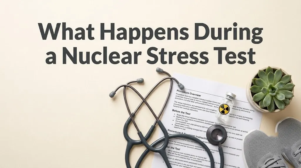
The stress phase begins after the resting images are reviewed and found adequate. If you are having an exercise stress test, you will walk on a treadmill while the speed and incline increase every three minutes according to a standardized protocol, usually the Bruce protocol. Your heart rate, blood pressure, ECG, and symptoms are monitored continuously. When you reach your target heart rate, typically 85 percent of your age-predicted maximum, or when you develop symptoms, ECG changes, or fatigue that prevents continuing, a second dose of tracer is injected through your IV. You then exercise for an additional minute to allow the tracer to circulate while your heart is still under stress. If you are receiving a chemical stress test instead, you will lie on a stretcher while a medication such as regadenoson, adenosine, or dobutamine is infused through your IV to dilate your coronary arteries or increase your heart rate pharmacologically, and the tracer is injected at the point of maximum pharmacologic effect.
The post-stress scan follows a waiting period of 30 to 45 minutes, during which you may be asked to drink water and eat a small snack to encourage tracer clearance from your liver and gallbladder, which sit adjacent to the heart and can interfere with image quality if not cleared. The second scan mirrors the first in duration and positioning. After both sets of images are captured, a nuclear medicine physician and your cardiologist will review the rest and stress perfusion maps side by side, looking for areas where blood flow appears normal at rest but diminished during stress. Those reversible perfusion defects indicate coronary arteries that are open enough to supply the heart at rest but too narrowed to meet the increased oxygen demand of exertion.
What to Expect Nuclear Stress Test
What you can expect during a nuclear stress test is that most of the experience will involve waiting quietly between phases rather than undergoing active procedures. The injection of the tracer feels like any standard IV injection, with a brief cool sensation in the arm but no burning, warmth, or unusual systemic effects. The tracer itself is colorless, odorless, and produces no physical sensations. You will not feel radioactive, and you will not notice the gamma camera detecting the tracer. The camera does not emit radiation. It only detects the radiation leaving your body from the tracer that has already been absorbed by your heart tissue.
The treadmill portion, if you are having exercise stress, will feel like a progressively more challenging workout. You will start walking slowly on a flat surface, and every three minutes the speed and incline will increase. The goal is to push your heart to a level of exertion where any blood flow limitations become visible on the perfusion images. You may feel short of breath, your legs may fatigue, and you will definitely notice your heart pounding in your chest. This is the intended physiological state. The test is designed to stress your cardiovascular system, and the monitoring team is trained to watch for signs that the stress level has gone far enough. If you experience chest discomfort, significant shortness of breath, or dizziness, you will tell the supervising cardiologist or nurse immediately. The test can be stopped at any point, and the monitoring team can respond instantly to any concerning changes on your ECG or blood pressure readings.
The chemical stress experience is different in character but similar in purpose. Instead of your legs generating the cardiac workload, a drug dilates your coronary blood vessels or directly stimulates your heart. With adenosine or regadenoson, you may feel flushing in your face and chest, a warm sensation spreading through your body, mild shortness of breath, or a brief sense of chest pressure that is not the same as angina but can feel uncomfortable. These sensations typically resolve within one to two minutes after the infusion stops because the half-life of these drugs is measured in seconds, not hours. The technologist will tell you in advance exactly what to expect, and the sensations, while sometimes intense, are brief and self-limited. You will be monitored continuously, and the supervising physician will be in the room or immediately available.
The period between scans is uneventful for most people. You will sit in a recliner or waiting area, and you may be given water or a light snack to help clear tracer from the abdominal organs. This waiting period is not downtime for the staff but an intentional physiological window that allows the tracer to redistribute and the non-cardiac background activity to diminish. After the second scan is complete, the technologist removes your IV, and you are free to leave unless your cardiologist has specified additional monitoring. The entire process from check-in to discharge averages three and a half hours, though some protocols using a one-day rest-stress sequence with technetium can be completed in approximately two and a half hours.
How to Prepare for Nuclear Stress Test
Preparing correctly for a nuclear stress test is the single most controllable factor in whether your test produces a diagnostically useful result or gets canceled and rescheduled. The preparation requirements differ depending on whether you are having an exercise-based test or a chemical stress test, and your cardiologist’s office will give you specific instructions. The most important preparation rule, particularly for chemical stress testing, involves caffeine. Caffeine blocks adenosine receptors, the same receptors that vasodilator drugs like adenosine and regadenoson are designed to activate. If caffeine is occupying those receptors when the drug is infused, your coronary arteries will not dilate properly, and the test will show a false-negative result that makes your heart look healthier than it actually is.
For a vasodilator chemical stress test, you must eliminate all caffeine for a full 12 to 24 hours before your appointment. This includes coffee, tea, both black and green, energy drinks, soda including diet varieties, chocolate in any form including dark chocolate and cocoa, and even decaffeinated coffee and tea, which still contain small amounts of caffeine that can interfere with the test. Many over-the-counter headache medications contain caffeine, and you must check the labels of anything you take in the 24 hours before your test. If you consume caffeine inadvertently and do not inform the testing staff, the test will go forward, the images will be acquired, and the result may be uninterpretable or misleading. You will have spent hours undergoing an expensive procedure that fails to answer the clinical question it was ordered to address.
Food and drink restrictions apply to all stress test types. You should not eat solid food for four to six hours before the test because a full stomach can cause abdominal discomfort during exercise and because eating diverts blood flow to the digestive system. Water is generally allowed and encouraged in moderation before the test, though you should confirm with your specific facility because policies vary slightly. If you are diabetic, your preparation instructions will include specific guidance about how to manage your blood sugar and whether to adjust your insulin or oral medications on the morning of the test. Failing to address this in advance can result in hypoglycemia during the fasting period or hyperglycemia that affects tracer distribution.
Medication management is the preparation domain where errors most commonly occur, and the consequences can include both canceled tests and misleading results. Beta blockers, including metoprolol, atenolol, and carvedilol, blunt the heart rate response to exercise and can prevent you from reaching the target heart rate required for a diagnostic exercise stress test. Your cardiologist will instruct you whether to hold these medications for 24 to 48 hours before the test. Calcium channel blockers, nitrates, and certain heart rate-lowering drugs may also need to be held. Do not make this decision independently. Your cardiologist must weigh the diagnostic benefit of holding the medication against the clinical risk of interrupting treatment, and that calculation is specific to your cardiac history and the reason the test is being ordered.
Wear comfortable, loose-fitting clothing and flat walking shoes with non-slip soles suitable for treadmill exercise. You will change into a hospital gown from the waist up for the imaging portions, but your lower body remains in your own clothing for the treadmill phase. Leave jewelry at home, and avoid applying lotion, powder, or deodorant to your chest area on the day of the test, as these can interfere with ECG electrode adhesion. Bring your insurance card, photo identification, and a list of your current medications including dosages and the time you last took each one. Arrange for someone to drive you home afterward if you are receiving a chemical stress agent, as the vasodilator drugs can cause transient dizziness or lightheadedness that makes driving inadvisable for a few hours.
Can You Drink Water Before a Nuclear Stress Test
You can drink water before a nuclear stress test, and in fact most nuclear cardiology labs encourage moderate water intake in the hours leading up to your appointment. Water does not interfere with the radioactive tracer, the vasodilator drugs used in chemical stress testing, or the cardiac monitoring equipment. Staying adequately hydrated makes your veins easier to access for the intravenous line, which reduces the discomfort of IV placement and the likelihood that the technologist will need to attempt multiple sites. Dehydration can make venous access more difficult and may cause a drop in blood pressure during the stress portion of the test, particularly if you are receiving vasodilator medications that already lower systemic vascular resistance.
The water allowance is not unlimited, and there is a distinction between small amounts of water and drinking so much that your stomach is uncomfortably full during the treadmill phase or while lying flat for imaging. A full bladder can also become uncomfortable during the extended imaging sessions, and you will not be able to get up to use the restroom while the gamma camera is acquiring images without disrupting the scan and potentially requiring a repeat acquisition. Sip water in the hours before your test rather than drinking a large volume right before walking into the lab. A reasonable guideline is to drink a normal amount of water with any morning medications you have been instructed to take, and then drink small additional amounts if you feel thirsty.
What you cannot drink is any liquid that contains caffeine, as discussed in the preparation section. This prohibition includes coffee, tea, cola, energy drinks, and some clear sodas that contain caffeine. If you are uncertain whether a beverage contains caffeine, check the label or avoid it entirely. The risk is a chemically invalidated test that shows normal perfusion when coronary disease actually exists. The same prohibition applies to alcohol, which should be avoided for 24 hours before the test because it can affect heart rate and blood pressure in ways that confound the stress portion. If your specific facility allows clear liquids other than water before the test, they will tell you during the pre-procedure instruction phone call. Do not assume that what applies to one hospital or imaging center applies to another. Ask directly and follow the instructions you are given.
For readers who take morning medications with water, confirm with your cardiologist’s office whether those specific medications should be taken or held on the morning of the test. Many cardiac medications should be taken as usual with a small sip of water, while others, particularly beta blockers and some calcium channel blockers, may need to be held to avoid blunting the heart rate response. The water you use to swallow your pills is not the concern. The medication inside the pill is what matters, and you need explicit, personalized instructions about which medications to take and which to skip.
Key Takeaway: Caffeine is the single most common reason a nuclear stress test gets rescheduled or produces a nondiagnostic result. Eliminate it completely for 24 hours before a chemical stress test, and when in doubt about whether a food or drink contains caffeine, assume it does and skip it.
What Do They Do in a Nuclear Stress Test
The clinical team performing your nuclear stress test includes a nuclear medicine technologist, a registered nurse, and a supervising physician who is either a cardiologist or a nuclear medicine specialist depending on the facility. The technologist manages the gamma camera, administers the tracer injections, and acquires the images that will be used for interpretation. The nurse places your IV, monitors your vital signs during the stress portion, and manages any symptoms that arise during the procedure. The supervising physician reviews the stress ECG in real time, makes the decision about when the stress level is adequate for tracer injection, and is immediately available if any concerning changes occur.
The technologist begins by explaining each step of the procedure, answering your questions, and positioning the ECG electrodes on your chest. These electrodes connect to a cardiac monitor that displays your heart rhythm continuously throughout the test. The resting scan is acquired first in the most common protocol, which is a one-day rest-stress sequence. You lie on a padded table with your left arm extended above your head to move the arm bones out of the imaging field. The gamma camera moves slowly around your chest in an arc, stopping at multiple positions to acquire images from standardized angles. You must lie still during acquisition because motion blurs the images and can create artifacts that look like perfusion defects. The technologist will tell you when you can move between acquisitions.
The stress portion follows the resting scan. If you are exercising, the technologist or nurse explains the treadmill protocol, shows you how to use the handrails for balance without gripping them tightly enough to create ECG artifact, and reminds you to communicate any symptoms immediately. The treadmill starts slowly and flat, and you will walk while the machine gradually increases speed and incline. Your blood pressure is measured every two to three minutes, and the ECG is watched continuously for changes. When you reach the target heart rate, or if you develop diagnostic ECG changes, significant symptoms, or fatigue that prevents continuing, the technologist injects the second dose of tracer through your IV. You then continue walking for one additional minute to let the tracer circulate while your heart remains stressed.
If you are receiving a chemical stress test, you will not walk on the treadmill. Instead, you lie on a stretcher or reclining chair while the vasodilator drug is infused through your IV. The nurse monitors your blood pressure, heart rate, and ECG continuously during the infusion. When the drug has achieved maximum vasodilation, typically within two to three minutes for regadenoson, the tracer is injected, and the infusion is stopped. The drug effects dissipate rapidly, and the monitoring continues for several minutes after the infusion ends to ensure your vital signs return to baseline. The post-stress scan follows a 30- to 60-minute waiting period during which you may drink water and eat a small snack to encourage clearance of tracer from the liver and gallbladder. The second set of images is acquired using the same positioning and camera movements as the first set, and the entire study is then processed into a color-coded perfusion map that the interpreting physician compares side by side.
How to Interpret Nuclear Stress Test Results
Nuclear stress test results are interpreted by comparing the resting perfusion images with the stress perfusion images across every segment of the left ventricle. A normal result shows uniform, bright uptake of the radioactive tracer throughout the heart muscle at rest and during stress. There are no areas where tracer uptake appears diminished in either set of images. This finding, described in the report as normal myocardial perfusion, indicates that blood flow to your heart muscle is adequate both at rest and under the increased demand of exercise or pharmacologic stress. According to the American Society of Nuclear Cardiology, a normal nuclear stress test carries an annual risk of a major cardiac event of less than 1 percent for the following one to two years in patients without high-risk clinical features.
An abnormal result falls into two main categories: reversible perfusion defects and fixed perfusion defects. A reversible defect appears as an area of reduced tracer uptake during the stress images that was not present or was less pronounced on the resting images. This pattern indicates myocardial ischemia: a coronary artery that is open enough to supply the heart at rest but too narrowed to meet the increased oxygen demands of exertion. The location and extent of the reversible defect tells your cardiologist which coronary artery is likely narrowed and how much heart muscle is at risk. A reversible defect in the anterior wall and septum suggests a left anterior descending artery stenosis. A defect in the lateral wall suggests a circumflex artery lesion. A defect in the inferior wall points toward the right coronary artery.
A fixed perfusion defect shows reduced tracer uptake that is present on both the rest and stress images. This pattern typically represents scar tissue from a prior myocardial infarction, though it can sometimes reflect hibernating myocardium: heart muscle that is alive but chronically underperfused and has downregulated its contractile function. Distinguishing between scar and hibernating tissue sometimes requires additional testing, such as a viability study with a different tracer like thallium-201 or a cardiac PET scan. Your cardiologist will correlate the nuclear scan findings with your clinical history, your ECG, and any prior cardiac imaging to determine whether the fixed defect represents old infarction or potentially recoverable tissue.
The report also includes quantitative data that goes beyond the visual interpretation of the images. The left ventricular ejection fraction, a measure of how much blood the heart pumps with each contraction, is calculated from the nuclear data. Transient ischemic dilation, where the left ventricle cavity appears larger on stress images than on rest images, is a high-risk finding that suggests severe, extensive coronary disease affecting multiple vessels. Increased tracer uptake in the lungs during the stress images indicates elevated left ventricular filling pressures and is another high-risk marker. Your cardiologist will explain these findings in the context of your overall clinical picture, and the results will guide decisions about medication adjustments, further testing such as coronary angiography, or referral for revascularization procedures.
The following table summarizes the main nuclear stress test result patterns and what they indicate:
| Result Pattern | Rest Image | Stress Image | Clinical Meaning |
|---|---|---|---|
| Normal Perfusion | Uniform tracer uptake | Uniform tracer uptake | No significant coronary artery disease |
| Reversible Defect | Normal or near-normal uptake | Reduced uptake in a specific region | Ischemia from a narrowed coronary artery |
| Fixed Defect | Reduced uptake | Reduced uptake, unchanged | Prior myocardial infarction or hibernating tissue |
| Transient Ischemic Dilation | Normal LV cavity size | LV cavity appears larger | High-risk extensive disease |
What to Expect After a Nuclear Stress Test
What you can expect after a nuclear stress test is that you will be able to resume most normal activities immediately, though there are a few specific precautions to follow for the remainder of the day. The radioactive tracer used in the test, technetium-99m, has a physical half-life of approximately six hours, meaning that half of the radioactivity decays every six hours. The tracer is eliminated from your body primarily through urine, and drinking extra fluids in the hours after the test accelerates this clearance. By the following morning, the amount of residual radioactivity in your body is negligible, and you can interact normally with family members, including children and pregnant women, without any special precautions beyond what the discharge instructions specify.
The IV site in your arm may be slightly tender or bruised, and you can apply a cold pack if it feels sore. The ECG electrode removal may leave small red marks on your chest that fade within a day. If you performed the exercise portion of the test, you may feel tired or notice mild muscle soreness in your legs similar to what you would feel after a brief vigorous workout. This is normal and resolves with rest and hydration. If you received a chemical stress agent, any flushing, warmth, shortness of breath, or chest pressure you experienced during the infusion should have resolved within minutes. If those sensations persist or recur after you leave the facility, you should contact your cardiologist’s office for guidance.
Your cardiologist will schedule a follow-up appointment, either in person or by phone, to discuss your results. The timing of this discussion varies by practice. Some cardiologists review the images immediately and discuss preliminary findings before you leave the facility. Others wait until the nuclear medicine physician has completed the formal interpretation, which may take 24 to 48 hours, and then communicate the results at a scheduled follow-up visit. You should ask before leaving the testing facility when and how you will receive your results so that you are not left wondering and waiting without a timeline.
There are activities you should avoid for the remainder of the test day. Do not drive yourself home if you received a chemical stress agent, as vasodilator drugs can cause delayed dizziness or lightheadedness. Do not engage in strenuous exercise or heavy lifting until the following day, both to let your body recover and to avoid any vasovagal episodes at the IV site. You can shower normally after the test, and you can eat and drink whatever you normally would, though avoiding caffeine for the remainder of the day is sensible if your test was a chemical study to ensure no residual caffeine effects confound anything. The small amount of radioactivity in your body is not a meaningful risk to others, but some facilities advise avoiding prolonged close contact with pregnant women and infants for the first six to eight hours after the test out of an abundance of caution. Your discharge instructions will specify if this applies.
Exercise Stress Versus Chemical Stress During the Nuclear Test
The choice between walking on a treadmill and receiving a chemical infusion during your nuclear stress test is not a preference question like choosing between coffee and tea. It is a clinical decision based on whether your body can safely achieve the target heart rate through physical exertion. Exercise stress is the preferred method whenever possible because it provides additional diagnostic information beyond what the perfusion images alone reveal. Your exercise capacity measured in metabolic equivalents, your heart rate response, your blood pressure response, and any symptoms or ECG changes that develop during exercise all contribute to risk stratification. A patient who walks for nine minutes on the Bruce protocol with a normal heart rate and blood pressure response has a better prognosis than a patient who walks for three minutes and develops significant ST-segment depression, even if both have normal perfusion images.
Chemical stress is indicated when you cannot exercise adequately. Common reasons include severe arthritis or orthopedic limitations that prevent walking on a treadmill, peripheral artery disease that causes leg pain with exertion, neurological conditions that impair balance or coordination, deconditioning from prolonged illness, or medications that prevent an adequate heart rate increase. If you have a left bundle branch block on your resting ECG, chemical stress with a vasodilator agent is preferred because exercise-induced heart rate increases can produce false-positive septal perfusion defects in people with this conduction abnormality. Your cardiologist determines the appropriate stress method when ordering the test, not you, and the decision is based on your specific clinical characteristics.
The vasodilator drugs used for chemical stress, primarily regadenoson and adenosine, work through a mechanism that is physiologically distinct from exercise. Instead of increasing myocardial oxygen demand by making the heart work harder, they dilate the coronary arteries directly, increasing blood flow through healthy vessels by three to five times baseline. Narrowed vessels cannot dilate proportionally, so the relative difference in perfusion between healthy and diseased territories becomes visible on the nuclear images. This is a different physiological stress than exercise, but it produces diagnostically equivalent perfusion information for most patients. Dobutamine is used when vasodilator agents are contraindicated, such as in patients with severe asthma or COPD where adenosine receptor activation could trigger bronchospasm. Dobutamine increases heart rate and contractility directly, mimicking exercise more closely.
Your experience during the test will differ based on which method is used, but the imaging protocol, the tracer administration, and the post-stress scan are essentially identical regardless of stress type. The critical preparation difference is the caffeine restriction for vasodilator chemical stress, which does not apply to dobutamine stress. If your test order says “nuclear stress test with regadenoson” or “adenosine nuclear stress,” the caffeine prohibition is absolute. If it says “exercise nuclear stress test” or “dobutamine stress,” caffeine is not the same concern, though you should still follow your specific preparation instructions regarding food and medications.
Key Takeaway: The stress method is a medical decision based on your physical capabilities and your ECG characteristics, not a personal preference option. If you can exercise adequately, you will. If you cannot, the chemical option produces equivalently diagnostic perfusion information through a different physiological mechanism.
Radiation Exposure, Safety, and What Happens to the Tracer
The radioactive tracer used in nuclear stress testing delivers a radiation dose that is roughly comparable to what you receive from natural background sources over one to two years, and it is eliminated from your body within a day. Technetium-99m sestamibi, the most commonly used agent, delivers an effective radiation dose of approximately 9 to 12 millisieverts for a complete rest-stress study using the one-day protocol. This is less than the annual occupational exposure limit for nuclear workers and substantially less than the radiation dose from a diagnostic coronary catheterization. The risk of radiation-induced malignancy from a single nuclear stress test is estimated to be extremely small, on the order of 1 in 10,000 or less, and must be weighed against the immediate clinical benefit of accurately diagnosing coronary artery disease that could cause a myocardial infarction if undetected.
The tracer does not remain in your body permanently. Technetium-99m decays away with a six-hour physical half-life and is cleared through the kidneys into your urine. Thallium-201, which is used less commonly and primarily for viability studies, has a longer half-life of 73 hours and a somewhat higher radiation dose per unit of administered activity. The imaging protocols used in 2026 are designed to use the lowest tracer dose that produces diagnostically adequate images, and modern gamma cameras with solid-state detectors have improved sensitivity that allows for dose reductions compared with older equipment. The American Society of Nuclear Cardiology’s imaging guidelines emphasize using the lowest radiation exposure that achieves the diagnostic objective.
Safety during the test extends beyond radiation considerations. The stress portion, whether exercise or chemical, is supervised by personnel trained to recognize and respond to complications immediately. The risk of a serious adverse event during exercise stress testing is approximately 1 in 2,500 to 1 in 10,000 tests. The risk during vasodilator chemical stress is similar, with serious arrhythmias or myocardial infarction occurring in fewer than 1 in 1,000 tests. The defibrillator, emergency medications, and resuscitation equipment are in the room or immediately adjacent. The supervising physician is present or immediately available throughout the stress phase. These safety measures are why the test is performed in a supervised medical setting rather than at home: the small but real risk of a serious complication is managed by the clinical team’s ability to respond instantly.
You should inform the technologist and the supervising physician if you have had a prior adverse reaction to a stress test, if you have a history of serious arrhythmia, if you have an implantable cardioverter-defibrillator, or if you have severe aortic stenosis. These conditions do not necessarily preclude testing but may require modification of the protocol or additional monitoring. Pregnant women should not undergo nuclear stress testing unless the clinical need is urgent and cannot wait until after delivery, because the tracer crosses the placenta and delivers radiation to the fetus. If you are breastfeeding, you will need to pump and discard breast milk for a specified period after the test, typically 12 to 24 hours for technetium-99m agents, and your facility will provide specific instructions.
What a Nuclear Stress Test Can and Cannot Tell Your Doctor
A nuclear stress test tells your cardiologist whether there are regions of your heart muscle that are not receiving adequate blood flow during stress, and it localizes those regions to specific coronary artery territories. It provides a quantitative estimate of left ventricular function and can identify high-risk features such as transient ischemic dilation or increased lung uptake. These findings directly inform decisions about whether you need coronary angiography, medication optimization, or revascularization. A normal nuclear stress test is a powerful predictor of low short-term cardiac risk, and for many patients, it provides the reassurance needed to avoid more invasive testing.
What a nuclear stress test cannot do is visualize the coronary artery walls directly. It shows the functional consequence of a blockage, the perfusion deficit downstream, rather than the blockage itself. A coronary artery could have moderate plaque that does not limit flow enough to produce a perfusion defect but could still rupture and cause an acute myocardial infarction. Nuclear stress testing is a test for flow-limiting stenoses, not for vulnerable plaque. This is why a normal nuclear stress test does not guarantee that you will not have a cardiac event. It means that your risk of a flow-limiting stenosis is low, which in turn correlates with lower near-term event risk, but atherosclerosis is a disease of the artery wall, and perfusion imaging assesses the downstream consequence rather than the wall itself.
The test also cannot distinguish between different causes of chest pain or shortness of breath that are not cardiac in origin. If your nuclear perfusion images are normal but you continue to experience symptoms, your cardiologist will consider non-cardiac causes such as esophageal spasm, costochondritis, pulmonary disease, or anxiety-related chest discomfort. The nuclear stress test answers the cardiac perfusion question with high accuracy. It does not answer the broader question of what is causing your symptoms if the cause lies outside the coronary circulation.
False positives and false negatives occur with nuclear stress testing, as with any diagnostic test. Breast tissue attenuation, diaphragmatic attenuation, and patient motion during image acquisition can create artifacts that look like perfusion defects. A skilled interpreting physician can often recognize these artifacts and recommend additional imaging or a different test modality if the findings are equivocal. Balanced ischemia, where all three coronary arteries are equally narrowed so that no regional difference in perfusion appears, can produce a falsely reassuring study because every segment of the heart is equally underperfused. This is a well-recognized limitation of relative perfusion imaging, and it is one reason your cardiologist considers the entire clinical picture, including your symptoms, exercise capacity, ECG response, and quantitative data, rather than looking only at the color-coded perfusion maps.
The Cost Implications of a Nuclear Stress Test in 2026
A nuclear stress test in 2026 will cost between $1,500 and $5,500 depending on your insurance coverage, the facility where it is performed, and whether chemical stress agents are required. For someone with commercial insurance, the patient responsibility typically falls between $200 and $1,200 after copays, deductibles, and coinsurance are applied. An uninsured patient paying cash can expect a self-pay rate of $1,600 to $3,200 at an independent imaging center, with hospital-based facilities charging substantially more due to facility fees that can add $1,200 to $2,400 to the total. You should call your insurance company with the specific CPT code, which is 78452 for a nuclear perfusion study, and ask for the contracted rate at the facility where your test is scheduled before you arrive.
The chemical stress portion adds cost because the pharmacologic agents are billed separately. Regadenoson (Lexiscan) typically costs the facility $200 to $400 per dose, and that cost passes through to your bill as a separate line item or is included in the facility fee depending on how the billing is structured. If you are having an exercise nuclear stress test, there is no drug cost for the stress portion. The imaging components, the tracer, the technologist’s time, and the professional interpretation fees are the same regardless of stress method. The total cost difference between an exercise nuclear test and a chemical nuclear test is typically $400 to $900, attributable to the drug and the additional monitoring time required during the infusion.
Your insurance plan’s prior authorization requirements are not trivial. Many insurers, including UnitedHealthcare, Aetna, and Anthem, now require the ordering cardiologist to submit documentation supporting the medical necessity of nuclear imaging rather than a standard exercise ECG or stress echocardiogram. If the authorization is not in place before your test, your claim may be denied, and you could be responsible for a substantial balance. Your cardiologist’s office handles the prior authorization process in most cases, but you should confirm that it has been completed before you arrive for the test. A denied claim that could have been prevented by a pre-authorization phone call is a costly administrative oversight.
The financial takeaway is that nuclear stress testing is an investment in diagnostic clarity that can prevent much more expensive downstream events. A heart attack that results in hospitalization and stent placement costs the healthcare system $20,000 to $30,000 or more. A nuclear stress test that identifies ischemia before it progresses to infarction, allowing for targeted medical therapy or elective revascularization, has saved that entire expenditure. The test’s cost must be understood in that context. It is expensive in absolute dollars, but it is considerably less expensive than the events it is designed to prevent.
Key Takeaway: The cost you actually pay for a nuclear stress test is determined by three variables: whether the facility is hospital-based or independent, whether your deductible has been met, and whether the test uses chemical or exercise stress. Knowing these three numbers before you schedule lets you make an informed decision about where to have the test performed.
Questions to Ask Your Cardiologist Before the Test
The time to ask your cardiologist questions about the nuclear stress test is during the office visit when the test is ordered, not when you are lying on the imaging table with an IV in your arm. The most important question to ask is why this specific test was chosen rather than a standard exercise stress test, a stress echocardiogram, or a cardiac CT angiogram. Each test answers a slightly different clinical question, and each has different radiation exposure, cost, and diagnostic accuracy profiles. If your cardiologist can explain clearly why nuclear imaging is the right choice for your specific situation, you will have more confidence going into the procedure and a better understanding of what the results will and will not tell you.
Ask what the plan is if the test is abnormal. A nuclear stress test that shows a moderate area of reversible ischemia will likely lead to a recommendation for coronary angiography. If you are not prepared to undergo angiography or are uncertain about the next step, have that conversation before the stress test, not after the results arrive. Understanding the clinical pathway that follows from each possible result helps you participate in decision-making rather than being carried along by a sequence of tests and procedures that you did not anticipate.
Ask about your medication instructions specifically by name. Do not accept “hold your heart medications” as adequate guidance. Say, “I take metoprolol 50 milligrams, lisinopril 10 milligrams, and atorvastatin 20 milligrams. Which of these should I take the morning of the test, and which should I hold?” Write down the answers. Ask what time you should take the last allowed dose before the test. If the instructions are complicated or if you take multiple medications, ask the office to give you a printed instruction sheet that you can reference the night before and the morning of the test. Medication errors are one of the most common reasons for canceled or nondiagnostic stress tests, and they are entirely preventable with clear, specific communication.
Ask who will interpret your results and how you will receive them. Some cardiologists read nuclear studies themselves. Others send the images to a nuclear medicine physician for formal interpretation and then incorporate those findings into their clinical assessment. Knowing who is reading your study and when you can expect to hear the results prevents the anxiety of waiting without a timeline. If your test is on a Thursday and the interpreting physician does not finalize reports over the weekend, you may not hear back until Monday or Tuesday. Knowing this in advance is better than spending the weekend checking your patient portal every two hours.
Frequently Asked Questions About What Happens During a Nuclear Stress Test
What happens during a nuclear stress test if I cannot walk on the treadmill?
If you cannot walk on a treadmill, you will receive a chemical stress test where a medication such as regadenoson or adenosine is infused through your IV to dilate your coronary arteries.
The drug simulates the increased blood flow that exercise would produce, and the tracer is injected at the point of maximum pharmacologic effect.
You will be monitored continuously during the infusion, and any side effects like flushing or mild chest pressure typically resolve within one to two minutes after the infusion stops.
What happens during nuclear stress test if I have a bad reaction?
Serious reactions during nuclear stress testing are rare, occurring in fewer than 1 in 1,000 tests, and the clinical team is trained to respond immediately with emergency medications, defibrillation, and physician intervention.
The supervising cardiologist is present or immediately available during the entire stress phase and monitors your ECG, blood pressure, and symptoms continuously.
If a concerning change occurs, the test is stopped, the appropriate intervention is delivered, and you will not leave the lab until you are stable.
Can I drink water before nuclear stress test if I take morning medications?
You can drink a small amount of water to swallow morning medications that your cardiologist has instructed you to take, and moderate water intake before the test is generally encouraged.
The water used to take your pills will not interfere with the tracer, the imaging, or the stress portion of the test.
Confirm with your cardiologist’s office which specific medications should be taken and which should be held, as the medication decision matters far more than the water used to swallow them.
How to interpret nuclear stress test results when they say reversible defect?
A reversible defect means the nuclear images show an area of reduced blood flow during stress that was not present at rest, indicating that a coronary artery is narrowed enough to limit blood flow when your heart is working hard.
The location of the defect tells your cardiologist which coronary artery is likely affected, and the size of the defect indicates how much heart muscle is at risk.
A reversible defect typically leads to further evaluation with coronary angiography or medication adjustments depending on the defect’s size, location, and your overall clinical picture.
What to expect after a nuclear stress test when results are normal?
A normal nuclear stress test means your heart muscle received adequate blood flow both at rest and during stress, indicating no flow-limiting coronary artery disease.
Your cardiologist will discuss the results and may shift the diagnostic focus to non-cardiac causes of your symptoms such as esophageal, pulmonary, or musculoskeletal conditions.
A normal study carries an annual cardiac event risk of less than 1 percent for the following one to two years in patients without high-risk clinical features.
How long does what they do in a nuclear stress test actually take?
The entire nuclear stress test process from check-in to discharge takes three to four hours, with most of that time spent waiting between the resting scan and the stress portion and then waiting again before the post-stress scan.
The actual imaging time is 15 to 25 minutes for each scan, and the stress portion on the treadmill or with chemical infusion takes 10 to 20 minutes.
You will be sitting or lying quietly for the majority of the appointment, so bringing something to read or do quietly during the waiting periods is advisable.
What Happens Next Depends on Your Results
The nuclear stress test is a tool that produces a specific, actionable answer about whether your heart muscle is getting enough blood. If the answer is yes, your cardiologist can confidently rule out flow-limiting coronary disease and focus on other explanations for your symptoms. If the answer is no, and the images show a reversible perfusion defect, you and your cardiologist now know the location, extent, and severity of the problem, and you can make decisions about medication, angiography, or revascularization based on concrete data rather than suspicion. Either way, you are better informed than you were before the test.
The preparation you do in the days before the test directly determines whether that answer arrives cleanly or gets muddied by caffeine, medications, or miscommunication. Eliminate caffeine completely if you are having vasodilator chemical stress. Get clear, written instructions about your medications. Ask your cardiologist the specific questions listed above before the appointment, not when you are on the table with an IV running. A nuclear stress test is a cooperative endeavor between you and the clinical team, and your half of the cooperation happens in the 24 hours before you walk through the door.
What you have learned here is the complete, phase-by-phase reality of what happens during a nuclear stress test, from check-in to discharge to results. You now know what the tracer does, why you wait between scans, what the treadmill feels like, how the chemical option works, and why caffeine is the enemy of an accurate vasodilator study. You also know what the test cannot tell your doctor and why a normal result does not mean you are immune to cardiac events. Walk into your appointment informed, prepared, and clear on the plan, and you will have done everything within your control to ensure the test delivers the answer it was ordered to provide.