What Is an Echo Stress Test in 2026: Complete Patient Guide
An echo stress test is a diagnostic procedure that combines ultrasound imaging of your heart with physical exercise or medication to evaluate how well your heart muscle pumps blood when it’s working harder than at rest. The test compares images of your heart before and immediately after stress to detect areas where blood flow may be reduced due to narrowed coronary arteries.
The American College of Cardiology and the American Heart Association include stress echocardiography in their guidelines as a first-line diagnostic test for evaluating patients with suspected coronary artery disease. According to the American Society of Echocardiography, stress echo has a sensitivity of approximately 80 to 85 percent and specificity of 84 to 86 percent for detecting physiologically significant coronary artery narrowing, making it one of the most reliable non-invasive cardiac tests available.
This article explains exactly what an echo stress test involves, how the exercise and medication versions differ, what the test can and cannot detect, how to prepare properly, and what your results mean. You’ll learn the cardiac physiology behind the test, not just the procedural steps, so you understand what your cardiologist is looking for and why.
What Is an Echo Stress Test
An echo stress test is a non-invasive cardiac diagnostic procedure that uses ultrasound to create images of your heart at rest and immediately after your heart rate has been elevated through exercise or medication. The test detects differences in how your heart muscle moves under stress compared to rest, revealing areas that may not be receiving adequate blood flow.
The term “echo” refers to echocardiography, which uses high-frequency sound waves produced by a handheld transducer placed on your chest. These sound waves bounce off heart structures and return to create real-time moving images of your heart chambers, valves, and muscle. The term “stress” refers to increasing your heart’s workload, either by having you exercise on a treadmill or stationary bicycle or by giving you medication that makes your heart work harder.
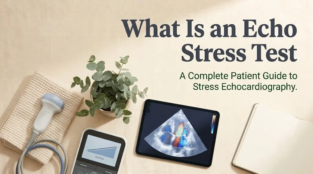
The fundamental diagnostic principle behind the test is the ischemic cascade. When a coronary artery is narrowed by atherosclerosis, blood flow to the heart muscle it supplies may be adequate at rest but insufficient when the heart works harder and demands more oxygen. This insufficient flow, called myocardial ischemia, causes the affected heart muscle to contract abnormally. The abnormal contraction, called a wall motion abnormality, appears on echocardiography before electrical changes appear on an ECG and before you would feel chest pain. This is why echo stress testing can detect coronary disease earlier and more accurately than exercise ECG alone.
The test evaluates the left ventricle, the heart’s main pumping chamber, in multiple cross-sectional views. A standard stress echo examines the left ventricle in several standard views, dividing it into segments according to the 17-segment model recommended by the American Society of Echocardiography. Each segment corresponds to the territory supplied by a specific coronary artery, allowing the cardiologist to localize which artery may be narrowed based on which wall segments show abnormal motion after stress.
What Is a Stress Echocardiogram
A stress echocardiogram is the formal medical name for an echo stress test. The terms are used interchangeably in clinical practice. “Echocardiogram” is the full term for an ultrasound examination of the heart. “Stress echocardiogram” specifies that the ultrasound is performed in conjunction with cardiac stress, either from exercise or medication.
The procedure has been in clinical use for over four decades and has a substantial evidence base supporting its diagnostic accuracy. The American Society of Echocardiography publishes and regularly updates practice guidelines for stress echocardiography, standardizing how the test is performed, interpreted, and reported across medical centers worldwide. This standardization is one of the test’s strengths. A stress echo performed according to guidelines in one hospital should yield comparable results to one performed in another.
A stress echocardiogram differs from a standard resting echocardiogram in its purpose and execution. A resting echo shows your heart’s structure and function under baseline conditions. It can detect valve problems, chamber enlargement, and resting wall motion abnormalities from prior heart attacks. A stress echo adds the dynamic component of comparing rest to stress, which is what reveals inducible ischemia, the hallmark of physiologically significant coronary artery disease. The two tests are complementary, and a stress echo always includes resting images as its baseline.
The stress echocardiogram also differs from other cardiac stress tests that do not include imaging. An exercise ECG stress test, sometimes called a regular stress test, monitors your heart’s electrical activity and blood pressure during exercise but does not take ultrasound images. Adding echocardiography significantly improves diagnostic accuracy. The images provide direct visual evidence of how the heart muscle is contracting, information that ECG alone cannot provide.
What Is a Stress Echocardiogram Test
A stress echocardiogram test is a complete diagnostic examination that includes resting echocardiography, a stress component to elevate your heart rate, and immediate post-stress echocardiography to capture images of your heart at peak workload. The entire test takes approximately 45 to 90 minutes from check-in to completion.
The test has three distinct phases. Phase one is the resting echocardiogram. You lie on an examination table while a sonographer places a transducer on several positions on your chest to obtain images of your heart at baseline. These images document your heart’s resting size, shape, and wall motion. They serve as the comparison for post-stress images.
Phase two is the stress component. If you are having an exercise stress echo, you will walk on a treadmill or pedal a stationary bicycle while your heart rate, blood pressure, and ECG are continuously monitored. The exercise intensity increases every three minutes according to a standardized protocol, typically the Bruce protocol on a treadmill. Your target is to reach at least 85 percent of your age-predicted maximum heart rate. If you are having a pharmacological stress echo, you will receive medication through an intravenous line while lying on the examination table. The medication, typically dobutamine, increases your heart rate and contractility without exercise.
Phase three is the post-stress echocardiogram. For exercise stress, you must move quickly from the treadmill to the examination table within 60 to 90 seconds after stopping exercise, because wall motion abnormalities from ischemia can resolve within minutes as your heart rate returns to baseline. The sonographer immediately begins acquiring the same views obtained at rest. For pharmacological stress, imaging continues throughout the medication infusion and recovery. A cardiologist later compares the rest and stress images side by side, evaluating each left ventricular segment for any change in wall motion or thickening.
Echo Stress Test
The echo stress test is one of several non-invasive cardiac stress tests available, and understanding how it compares to alternatives helps explain why a cardiologist might choose it for a particular patient. The main alternatives are the exercise ECG stress test without imaging and the nuclear stress test with myocardial perfusion imaging.
An exercise ECG stress test monitors heart rhythm and blood pressure during exercise but provides no direct images of the heart muscle. It is appropriate for patients with a normal resting ECG who can exercise adequately and have a low to intermediate pre-test probability of coronary disease. The addition of echocardiography provides greater diagnostic accuracy, particularly in patients with abnormal resting ECGs, women, and patients with intermediate risk.
A nuclear stress test uses a radioactive tracer injected during stress to create images of blood flow to the heart muscle. It provides perfusion information that echocardiography does not directly show, but it involves radiation exposure and takes longer. Nuclear imaging has similar sensitivity to stress echo for detecting coronary disease but may have slightly lower specificity in some populations. The choice between stress echo and nuclear stress often depends on local expertise, patient characteristics, and the specific clinical question being asked.
Stress echo has several advantages that make it a preferred first-line test in many situations. It uses no ionizing radiation, which is particularly relevant for younger patients and women of childbearing age. It provides structural information about heart valves, chamber sizes, and wall thickness that nuclear testing does not. It can detect non-coronary causes of symptoms including valvular heart disease. The primary limitation of stress echo is that image quality depends on the patient’s body habitus. In patients with obesity, lung disease, or chest wall abnormalities, ultrasound images may be suboptimal, and contrast agents or alternative testing may be needed.
| Test | What It Shows | Radiation | Duration | Best For |
|---|---|---|---|---|
| Exercise ECG only | Electrical activity, heart rate, blood pressure | None | 30 to 45 minutes | Low-risk patients, normal resting ECG |
| Stress echocardiogram | Wall motion, valve function, chamber size | None | 45 to 90 minutes | Most patients, women, abnormal resting ECG |
| Nuclear stress test | Myocardial perfusion | Yes | 2 to 4 hours | Patients with poor echo windows, specific perfusion questions |
Key Takeaway: An echo stress test combines rest and stress ultrasound images to detect wall motion abnormalities that indicate reduced blood flow from narrowed coronary arteries, providing more diagnostic information than exercise ECG alone without the radiation of nuclear testing.
Stress Echo Test
A stress echo test is ordered when a cardiologist or primary care physician needs to evaluate whether your symptoms are caused by coronary artery disease, assess the severity of known coronary disease, or evaluate your heart’s functional capacity before surgery or after a heart attack.
The most common reason for ordering a stress echo is to evaluate chest pain or chest discomfort suggestive of angina. Not all chest pain is cardiac, and a normal stress echo can provide strong reassurance that the symptoms are not from coronary disease. The test is also ordered for shortness of breath, particularly exertional dyspnea, which can be an angina equivalent, meaning reduced blood flow manifests as breathlessness rather than chest pain.
Stress echo is used to assess the functional significance of known coronary blockages. A coronary angiogram or CT scan may show anatomical narrowing, but not every narrowing restricts blood flow enough to cause ischemia. A stress echo can determine whether a particular blockage is physiologically significant by showing whether the heart muscle it supplies develops a wall motion abnormality under stress. This information guides decisions about whether a blockage needs stenting or bypass surgery or can be managed medically.
Pre-operative risk assessment is another common indication. Before major non-cardiac surgery, particularly vascular surgery, a stress echo can evaluate whether the heart can tolerate the increased demands of surgery and recovery. A normal stress echo at adequate workload is associated with low perioperative cardiac risk.
Stress echo is also used after a heart attack to assess residual ischemia and left ventricular function. The test can identify whether there is additional heart muscle at risk beyond the area already damaged, which guides decisions about further revascularization.
Cardiac Stress Echo
Cardiac stress echo is a specialized application of echocardiography performed by cardiologists and cardiac sonographers with specific training in stress imaging. The quality and diagnostic accuracy of the test depend heavily on the expertise of the team performing and interpreting it.
The sonographer is responsible for acquiring high-quality images at rest and immediately after stress. This requires technical skill with the ultrasound equipment and the ability to obtain standardized views rapidly. During exercise stress echo, the sonographer has only 60 to 90 seconds after exercise cessation to capture all required views before the heart rate drops and any wall motion abnormalities resolve. This time pressure makes stress echo technically more demanding than resting echocardiography.
The interpreting cardiologist evaluates the rest and stress images side by side, comparing each myocardial segment frame by frame. The assessment uses a standardized wall motion scoring system. Each of the 17 left ventricular segments is scored as 1 for normal, 2 for hypokinetic meaning reduced contraction, 3 for akinetic meaning absent contraction, or 4 for dyskinetic meaning outward bulging during contraction. A segment that is normal at rest but becomes hypokinetic or akinetic after stress indicates inducible ischemia in the territory of the coronary artery supplying that segment.
The cardiologist synthesizes the wall motion findings with the ECG response, heart rate achieved, blood pressure response, and any symptoms reported during stress to produce an integrated interpretation. The final report typically states whether the test was normal or abnormal, whether there is evidence of inducible ischemia, and if so, which coronary territory is likely involved and the extent of myocardium at risk.
Echocardiogram Stress Test
An echocardiogram stress test is the same procedure as a stress echocardiogram, with the word order simply reversed. This terminology variation can cause confusion, but all terms refer to the same diagnostic test combining cardiac ultrasound with physical or pharmacological stress.
The test evaluates several aspects of cardiac function beyond wall motion. It assesses left ventricular ejection fraction, the percentage of blood pumped out with each heartbeat, both at rest and after stress. In a normal heart, ejection fraction increases with exercise. A failure to increase, or a decrease, suggests significant coronary disease or cardiomyopathy. The test also evaluates heart valve function under stress conditions, which can reveal valve problems that are not apparent at rest.
Global left ventricular function provides prognostic information independent of regional wall motion. A patient with normal wall motion but a blunted ejection fraction response to exercise has a worse prognosis than someone whose ejection fraction increases appropriately, even if neither has a discrete wall motion abnormality. This global assessment adds value to the regional wall motion analysis.
Right ventricular function is also evaluated, though less extensively than the left ventricle. The right ventricle can show stress-induced dysfunction in certain conditions, including severe left coronary disease affecting the septum and primary pulmonary conditions. Right ventricular assessment during stress echo is less standardized than left ventricular assessment but can provide additional diagnostic information.
Key Takeaway: The cardiologist evaluates not just whether a wall moves abnormally, but how much myocardium is affected, how severe the abnormality is, which coronary artery territory is involved, and how global heart function responds to stress.
Does Echo Stress Test Show Blockage
An echo stress test does not directly show anatomical blockages in the way a coronary angiogram or CT coronary angiogram does. It shows the functional consequence of a blockage by detecting abnormal heart muscle contraction in areas that are not receiving adequate blood flow during stress.
The distinction between anatomical and functional testing is fundamental to understanding what a stress echo can and cannot tell you. An angiogram shows the coronary arteries directly, revealing the location and severity of narrowings. A stress echo shows whether those narrowings, or narrowings not yet imaged, are restricting blood flow enough to affect heart muscle function. A blockage that narrows a coronary artery by 50 percent may not cause ischemia and would not produce an abnormal stress echo. A blockage of 70 percent or greater is more likely to cause ischemia and produce a wall motion abnormality. The stress echo detects physiologically significant disease, the kind that matters for symptoms and prognosis.
The test has known sensitivity and specificity for detecting significant coronary artery disease. Sensitivity of 80 to 85 percent means the test will be abnormal in 80 to 85 percent of patients who have physiologically significant blockages. Specificity of 84 to 86 percent means the test will be normal in 84 to 86 percent of patients who do not have such blockages. These figures mean the test is very good but not perfect. False negatives, where significant disease is present but the test is normal, can occur if the patient does not achieve an adequate heart rate, if the blockage is in a location difficult to image, or if single-vessel disease affects only a small territory. False positives, where the test is abnormal but no significant disease is present, can occur in patients with baseline ECG abnormalities, left bundle branch block, or cardiomyopathy.
The test also cannot directly visualize non-obstructive plaque. Atherosclerosis begins long before it narrows an artery enough to restrict flow. A normal stress echo means there is no flow-limiting disease at the time of the test. It does not mean the coronary arteries are free of atherosclerosis. This is why risk factor modification remains important even with a normal stress echo result.
Stress Echocardiography
Stress echocardiography has evolved significantly since its introduction in the 1980s, with advances in ultrasound technology, contrast agents, and strain imaging that have improved both image quality and diagnostic accuracy.
Modern stress echo systems use harmonic imaging and digital acquisition that produce substantially better image quality than earlier equipment. Digital storage allows side-by-side comparison of rest and stress images in a continuous loop format that makes subtle wall motion differences easier to detect. These technical improvements have contributed to the test’s high diagnostic accuracy.
Ultrasound contrast agents can be used during stress echo when image quality is suboptimal. These agents consist of microscopic gas-filled microbubbles that are injected intravenously and opacify the left ventricular cavity, improving the definition of the endocardial border, the inner lining of the heart muscle. Better border definition allows more confident assessment of wall motion and thickening. Contrast agents are particularly helpful in patients with obesity, lung disease, or prior chest surgery where standard imaging is challenging.
Strain imaging is an advanced echocardiographic technique that quantifies myocardial deformation, providing an objective measure of contraction beyond the qualitative visual assessment of wall motion. Strain measures how much the heart muscle shortens during contraction. Reduced strain after stress can indicate ischemia even when visual wall motion assessment appears normal. Global longitudinal strain is the most commonly used strain parameter in stress echo and has been shown to improve diagnostic accuracy when added to standard wall motion analysis, according to research published in the Journal of the American College of Cardiology.
Myocardial contrast echocardiography, using the same contrast agents, can assess myocardial perfusion in addition to wall motion. This technique visualizes the entry of contrast into the heart muscle itself, showing areas of reduced blood flow directly. Perfusion imaging adds another dimension to stress echo that brings its diagnostic capabilities closer to those of nuclear perfusion imaging, without the radiation exposure.
Echo Stress Test Procedure
The echo stress test procedure follows a structured sequence designed to maximize diagnostic accuracy while minimizing patient discomfort and risk. Understanding each step reduces anxiety and helps you cooperate effectively with the testing team.
Upon arrival, you will be asked to change into a hospital gown and lie on an examination table. A cardiac sonographer will place adhesive electrode patches on your chest to monitor your ECG throughout the test. A blood pressure cuff will be placed on your arm. An intravenous line may be placed in a vein in your arm or hand, particularly if you are having a pharmacological stress test or if contrast agent might be needed.
The resting echocardiogram is performed first. The sonographer will apply a clear gel to several positions on your chest and will press a handheld transducer against your skin to obtain images. You may be asked to briefly hold your breath or shift position to optimize image quality. The transducer does not cause pain, though the pressure may be mildly uncomfortable. The resting images typically take 10 to 15 minutes to acquire all standard views.
The stress component follows immediately. For an exercise test, you will be positioned on a treadmill or stationary bicycle and instructed to begin exercising. The treadmill protocol starts slowly and increases speed and incline every three minutes. The exercise continues until you reach your target heart rate, develop symptoms that require stopping, or reach the maximum effort you can sustain. For a pharmacological test, the medication infusion begins while you remain on the examination table. Your heart rate, blood pressure, and ECG are monitored continuously.
The post-stress imaging is the most time-critical phase. For exercise stress, you must move from the treadmill to the examination table within 60 to 90 seconds. The sonographer immediately begins acquiring the same views obtained at rest, working quickly to capture images before your heart rate slows. For pharmacological stress, imaging occurs during the peak effect of the medication. You will be monitored for a recovery period until your heart rate and blood pressure return to baseline.
Exercise Stress Echo
Exercise stress echo uses physical activity, most commonly treadmill walking or stationary cycling, to increase the heart’s workload. This is the preferred method whenever possible because it uses the body’s natural physiological response to exercise rather than medication.
The treadmill protocol most commonly used is the Bruce protocol, which consists of seven three-minute stages of progressively increasing speed and incline. Stage one is walking at 1.7 miles per hour at a 10 percent grade. Each subsequent stage increases both speed and incline. The modified Bruce protocol starts at a lower workload for older or deconditioned patients. The goal is to reach at least 85 percent of your age-predicted maximum heart rate, calculated as 220 minus your age. Achieving this target is important because the sensitivity of the test for detecting ischemia depends on how close you get to your maximum heart rate.
Your physiological response to exercise provides diagnostic information beyond the echocardiographic images. The heart rate achieved, the blood pressure response, the rate-pressure product which is heart rate multiplied by systolic blood pressure and reflects myocardial oxygen demand, and any symptoms or ECG changes all contribute to the test interpretation. A normal blood pressure response is a progressive rise in systolic pressure with exercise. A drop in blood pressure during exercise is an abnormal response that can indicate severe coronary disease.
Exercise stress echo has advantages over pharmacological stress. It provides information about functional capacity and exercise tolerance that pharmacological testing cannot. It reproduces the conditions under which the patient’s symptoms typically occur. It avoids the side effects and risks of stress-inducing medications. The primary limitation is that not all patients can exercise adequately. Orthopedic limitations, peripheral vascular disease, neurological conditions, severe lung disease, and general deconditioning can prevent a patient from achieving the target heart rate through exercise.
Dobutamine Stress Echo
Dobutamine stress echo uses medication instead of exercise to increase the heart’s workload. It is indicated for patients who cannot exercise adequately due to physical limitations or for whom exercise testing is contraindicated.
Dobutamine is a medication that stimulates beta-adrenergic receptors in the heart, mimicking the effects of the sympathetic nervous system’s natural transmitters epinephrine and norepinephrine. At low doses, dobutamine increases the force of heart muscle contraction, called its inotropic effect. At higher doses, it also increases heart rate, called its chronotropic effect. The combination of increased heart rate and increased contractility raises myocardial oxygen demand, creating the same stress on the heart that exercise would produce.
The dobutamine infusion follows a standardized protocol. The medication is delivered through an intravenous line at progressively increasing doses, typically starting at 5 or 10 micrograms per kilogram per minute and increasing at three-minute intervals to a maximum of 40 or 50 micrograms per kilogram per minute. If the target heart rate is not achieved with dobutamine alone, small doses of atropine may be added to further increase heart rate. Echocardiographic images are obtained at baseline, at low dose, at peak dose, and during recovery.
An alternative to dobutamine is vasodilator stress using medications such as dipyridamole, adenosine, or regadenoson. These work through a different mechanism than dobutamine. Rather than increasing the heart’s oxygen demand, they dilate normal coronary arteries more than diseased ones, creating a steal phenomenon where blood flow is redirected away from territories supplied by narrowed arteries. Vasodilator stress is less commonly used for echocardiography than for nuclear perfusion imaging, but it is an option for selected patients.
Echo Treadmill Test
An echo treadmill test is the most common form of exercise stress echocardiography in the United States. The treadmill is preferred over the stationary bicycle in many centers because most patients are familiar with walking and can achieve higher workloads on a treadmill than on a bicycle.
The treadmill portion uses a motorized belt that increases in speed and incline according to the Bruce or modified Bruce protocol. You will be shown how to walk on the treadmill before the test begins. Handrails are available for balance but you should avoid gripping them tightly, as this reduces the workload on your heart. The test team will instruct you on proper treadmill walking technique.
Throughout the treadmill exercise, your ECG, heart rate, and blood pressure are displayed on monitors visible to the supervising clinician. You will be asked to report any symptoms including chest discomfort, shortness of breath, leg fatigue, dizziness, or palpitations. The test team watches for ECG changes, blood pressure abnormalities, and concerning symptoms that would indicate the test should be stopped.
Stopping criteria for the treadmill portion include reaching the target heart rate, developing severe chest pain or anginal equivalent, significant ECG changes such as ST-segment depression, a drop in systolic blood pressure, dangerous arrhythmias, or patient request due to fatigue or other symptoms. When the test is stopped, you must move quickly to the examination table, as described earlier. The ability to transition rapidly from treadmill to table and obtain images within the narrow post-exercise window is a key skill of the stress echo team.
What to Do Before a Stress Echo Test
What you do before a stress echo test can affect both the safety and accuracy of the procedure. Proper preparation ensures the test can be performed as planned and yields reliable results. The specific instructions vary somewhat between exercise and pharmacological stress echo and depend on your medical history.
Medication management is the most important preparation consideration. You will receive specific instructions about which medications to take and which to hold before the test. Beta-blockers, some calcium channel blockers, and certain heart rate-controlling medications are often held for 24 to 48 hours before an exercise stress echo because they prevent the heart rate from reaching the target level, reducing test sensitivity. Never stop medications on your own. Your ordering physician will provide written instructions about exactly which medications to adjust and which to continue.
Fasting instructions typically include nothing to eat or drink except small sips of water for three to four hours before the test. A full stomach can cause discomfort during exercise. Caffeine must be avoided for 12 to 24 hours before the test, particularly for pharmacological stress testing, because caffeine can interfere with the action of vasodilator medications used in some stress protocols.
Clothing and personal preparation contribute to a smooth test. Wear comfortable clothing and walking shoes suitable for exercise. Avoid dresses, skirts, or restrictive clothing. Do not apply lotions, creams, or oils to your chest area, as these can interfere with electrode adhesion and ultrasound gel contact. If you use a bronchodilator inhaler, bring it with you in case it is needed during or after exercise.
Transportation planning is sensible because you may feel tired after the test, particularly if you achieved a high exercise workload. While most people can drive themselves home after an uncomplicated stress echo, arranging for someone to drive you can reduce stress about the appointment. If you are having a pharmacological stress test with dobutamine, some centers recommend against driving immediately after.
Echo Stress Test Preparation
Echo stress test preparation involves several specific steps that optimize test conditions and minimize the chance of cancellation or inaccurate results. Taking these steps seriously improves the likelihood that your test will be diagnostic and conclusive.
Review your written preparation instructions as soon as you receive them. Instructions from different medical centers may vary. If you have questions about whether to take specific medications, call the testing center for clarification rather than guessing. Bring a list of your current medications with doses and timing to the test. The testing team needs to know exactly what you have taken and when.
Inform the testing team of relevant medical conditions during scheduling and upon arrival. Pregnancy, known or suspected, is a relative contraindication to pharmacological stress agents, though exercise stress echo with appropriate monitoring may be performed if clinically necessary. Severe aortic stenosis, uncontrolled hypertension, acute illness, and recent stroke or heart attack are potential contraindications that the ordering physician should have considered but that the testing team will verify.
The IV line placement for pharmacological testing or contrast administration is typically done in a forearm or hand vein. If you have a history of difficult IV access, mention this during scheduling so an experienced provider can be available. For exercise testing, IV access may or may not be needed depending on the protocol.
Managing pre-test anxiety is appropriate and common. Many patients feel nervous about a heart test, particularly if they have experienced cardiac symptoms. The testing team is experienced in working with anxious patients. Tell the sonographer or nurse if you’re feeling nervous. They can explain each step as it happens and answer questions. The test itself is closely monitored by medical professionals trained to recognize and respond to any problems.
Key Takeaway: Medication management per your physician’s specific instructions, fasting, avoiding caffeine and chest lotions, wearing exercise clothing, and reviewing instructions in advance are the preparation steps that most affect test safety and accuracy.
Stress Echo Results Explained
Stress echo results are interpreted by a cardiologist who compares the rest and stress images and integrates the imaging findings with the ECG, blood pressure, and symptom data collected during the test. Results are typically available within a few days, though preliminary findings may be discussed immediately after the test in some centers.
A normal stress echo result means that all left ventricular wall segments contracted normally at rest and showed normal or increased contraction after stress. The left ventricular ejection fraction increased appropriately with exercise. No wall motion abnormalities developed. The ECG and blood pressure responses were normal. This result indicates a low probability of physiologically significant coronary artery disease and is associated with a favorable prognosis. The annual risk of major cardiac events after a normal stress echo is less than 1 percent in appropriately selected patients.
An abnormal stress echo result shows one or more wall motion abnormalities that develop or worsen after stress compared to rest. The cardiologist describes which left ventricular segments are affected, the severity of the abnormality, and the coronary artery territory likely involved. A wall motion abnormality in the anterior wall and septum suggests disease in the left anterior descending artery. An abnormality in the inferior wall suggests right coronary artery disease. The lateral wall is typically supplied by the circumflex artery. The extent and severity of the abnormality correlate with the amount of myocardium at risk and guide decisions about further testing or treatment.
A non-diagnostic or equivocal result occurs when the test cannot definitively exclude or confirm ischemia. This may happen if the target heart rate was not achieved, if image quality was suboptimal, or if the findings are borderline. Your physician may recommend repeating the test with an alternative method, performing a different type of stress test, or proceeding to coronary angiography depending on your pre-test probability and clinical situation.
The cardiologist’s report also comments on non-coronary findings. Valve function at rest and during stress, chamber sizes, estimated pulmonary artery pressures, and any incidental findings are included. These provide a comprehensive cardiac assessment beyond the specific question of ischemia.
| Result | What It Means | Typical Next Step |
|---|---|---|
| Normal | No wall motion abnormality, good exercise capacity | Reassurance, risk factor management |
| Abnormal with ischemia | Wall motion abnormality develops with stress | May need angiography, medical therapy adjustment |
| Fixed wall motion abnormality | Scar from prior heart attack | Medical management, possibly viability assessment |
| Non-diagnostic | Target heart rate not achieved, poor images | Repeat with alternative method, consider angiography |
| Abnormal blood pressure response | Possible severe coronary disease or cardiomyopathy | Further evaluation, typically angiography |
Frequently Asked Questions About Echo Stress Testing
What is the difference between a regular stress test and an echo stress test?
A regular stress test monitors your ECG and blood pressure during exercise but does not take ultrasound images of your heart.
An echo stress test adds echocardiographic imaging before and after stress, providing direct visualization of heart muscle contraction that improves diagnostic accuracy substantially.
The echo version detects wall motion abnormalities that can reveal ischemia before ECG changes appear.
How long does an echo stress test take from start to finish?
An echo stress test takes approximately 45 to 90 minutes from check-in to completion.
The resting images take 10 to 15 minutes, the exercise or medication stress phase takes 10 to 20 minutes, and the post-stress imaging and recovery monitoring take another 15 to 20 minutes.
Additional time is needed for IV placement, electrode application, and pre-test verification if you are having a pharmacological stress test.
Can an echo stress test show blocked arteries?
An echo stress test shows the functional effect of blocked arteries by detecting abnormal heart wall motion in areas receiving insufficient blood flow during stress.
It does not directly visualize the coronary arteries the way a coronary angiogram or CT angiogram does.
A stress echo detects blockages that are severe enough to restrict blood flow during increased demand, which are the blockages that cause symptoms and pose risk.
Why would a doctor order an echo stress test?
A doctor orders an echo stress test to evaluate whether chest pain, shortness of breath, or other symptoms are caused by coronary artery disease.
The test is also ordered to assess the functional significance of known blockages, evaluate heart function before surgery, or determine prognosis after a heart attack.
A normal result can provide strong reassurance that symptoms are not cardiac in origin.
What happens if my echo stress test is abnormal?
An abnormal echo stress test showing inducible ischemia typically leads to further evaluation of your coronary arteries.
Your cardiologist may recommend coronary angiography to directly visualize the blockages and determine whether stenting or bypass surgery is needed.
Mild abnormalities may be managed with intensification of medical therapy and risk factor modification without immediate invasive testing.
Is an echo stress test painful or dangerous?
An echo stress test is not painful, though the exercise portion requires physical effort that may cause temporary fatigue, and the transducer pressure on your chest may be mildly uncomfortable.
Serious complications are rare, occurring in approximately 1 in 2,000 to 1 in 5,000 tests, and the testing team is trained to recognize and manage any problems immediately.
The diagnostic benefit of detecting significant coronary disease far outweighs the small procedural risk for appropriately selected patients.
An echo stress test gives your cardiologist something no symptom description or resting ECG can provide: a direct view of how your heart muscle responds when it’s working hard. The test detects the functional consequence of narrowed coronary arteries by revealing wall motion abnormalities that appear when blood flow cannot meet the heart’s increased demand. This physiological principle is why the test has remained a cornerstone of cardiac diagnosis for decades.
If you’re scheduled for this test, the most useful thing you can do is follow your medication instructions precisely and bring your medication list to the appointment. Wear comfortable walking shoes and a two-piece outfit that allows easy access to your chest. Tell the team if you’re nervous. They do this every day and can talk you through each step. The information your test provides could either give you powerful reassurance or identify a problem before it progresses to a heart attack.
Your cardiologist will explain your results in the context of your symptoms, risk factors, and overall health. A normal stress echo at an adequate workload is genuinely reassuring. An abnormal result gives you and your doctor actionable information about what needs attention and how to proceed. Either way, you’ll know more about your heart health after the test than before it, and that knowledge is the point.