What Is a PET Stress Test? 2026 Procedure & Meaning Guide
A PET stress test, short for Positron Emission Tomography stress test, is an advanced cardiac imaging procedure that evaluates how well blood flows through the arteries of your heart muscle during a state of induced “stress,” which is not psychological anxiety but a medically controlled simulation of physical exercise using a drug. If you have been told you need a “stress test” and immediately felt your heart race with worry, you have just experienced the exact semantic confusion this guide is designed to clear up. The “stress” in a PET stress test refers to the physiological workload placed on your heart, not your mental state.
The American Heart Association and the American College of Cardiology recognize PET stress testing as the gold standard for non-invasive myocardial perfusion imaging. It detects coronary artery disease with a higher degree of accuracy and with a lower radiation dose to the patient than the older, more common SPECT nuclear stress test. A 2023 meta-analysis published in the Journal of the American College of Cardiology confirmed that PET perfusion imaging has a sensitivity of approximately 90 percent and a specificity of 89 percent for detecting significant coronary blockages.
This guide will first clarify the critical distinction between a cardiac PET stress test and the imaging of psychological stress in the brain. Then, you will learn exactly how the pharmacological stress agents work at the receptor level, walk through the procedure step-by-step, and understand what your results mean. If you arrived at this article because you are feeling overwhelmed, anxious, and your heart is pounding, there is a section at the end that connects the dots between chronic psychological stress and heart health.
If you are in crisis or experiencing thoughts of self-harm, contact the 988 Suicide and Crisis Lifeline by calling or texting 988 at any time. This service is free, confidential, and available 24 hours a day.
what is a pet stress test
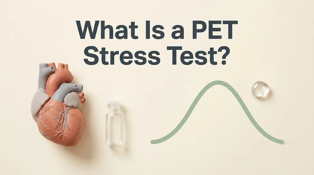
A PET stress test is a medical imaging procedure that uses a short-lived radioactive tracer and a PET scanner to create high-resolution, three-dimensional images of blood flow to the heart muscle at rest and during a state of pharmacological stress, which simulates the cardiovascular effect of exercise without requiring the patient to physically exert themselves. The test’s primary purpose is to detect areas of the heart that are not receiving adequate blood supply due to narrowed or blocked coronary arteries. This condition is called ischemia.
The acronym PET stands for Positron Emission Tomography. The scanner detects pairs of gamma rays emitted when a positron, released by the injected tracer, collides with an electron in the body. This annihilation event allows the scanner to precisely map where the tracer has accumulated. The tracers used for cardiac perfusion imaging are specifically designed to be taken up by healthy heart muscle cells in direct proportion to the blood flow they receive. Areas with low tracer uptake indicate low blood flow.
The “stress” component of the test name does not refer to psychological or emotional stress. This is the single most common point of confusion and anxiety for patients. A medical stress test physically stresses the cardiovascular system. This can be achieved through treadmill exercise, but in a PET stress test, it is most commonly achieved using a medication called a vasodilator. Vasodilators like adenosine or regadenoson temporarily widen the coronary arteries, increasing blood flow by 4 to 5 times the resting rate. This hyperemic response mimics what happens when you run on a treadmill.
A resting set of images is also acquired, either before or after the stress images, to provide a baseline. The cardiologist then compares the resting and stress images. If a region of the heart shows normal tracer uptake at rest but significantly reduced uptake during stress, the diagnosis is stress-induced ischemia. The artery supplying that region is likely blocked. If the uptake is poor at both rest and stress, the tissue is likely a scar from a previous heart attack.
Key Takeaway: The “stress” in a PET stress test is chemically-induced physical workload on your heart, not a psychological anxiety attack. The test itself will not stress you out emotionally if you know what to expect.
what is a pet scan stress test
A PET scan stress test is the identical procedure to a PET stress test, where the term “scan” is added to emphasize the imaging technology used. It refers to the combination of a PET scanner detecting the radioactive tracer and a pharmacological stress agent administered intravenously to evaluate myocardial perfusion. The terms “PET stress test,” “PET scan stress test,” and “cardiac PET perfusion study” are used interchangeably in clinical settings.
The critical clarification this query demands is that a PET scan is a tool, and it can be used to image many different physiological processes depending on the tracer injected. A cardiac PET stress test uses a perfusion tracer like Rubidium-82 or N-13 ammonia. These tracers are specifically formulated to map blood flow to the heart. However, the same PET scanner could be used with a completely different tracer, like Fluorodeoxyglucose (FDG) , to map glucose metabolism in a tumor or, critically for our niche, in the brain.
This is where the ambiguity peaks. A person experiencing severe psychological stress might wonder if a PET scan can “see” their anxiety. The answer is yes, but not as a routine clinical test. Research-grade PET scans using FDG or specialized tracers for neurotransmitter systems can absolutely visualize the neurobiological footprint of chronic stress. A 2022 study published in Psychoneuroendocrinology used FDG-PET to demonstrate that individuals with high allostatic load show increased metabolic activity in the amygdala, the brain’s fear center, and decreased activity in the prefrontal cortex, the area responsible for rational regulation of emotion.
So, a “PET scan stress test” can mean two distinct things. It most commonly means a cardiac perfusion test. Less commonly, in a research or specialized clinical context, it can mean a PET scan of the brain that measures the metabolic consequences of chronic psychological stress. This article focuses on the cardiac test, which is what your cardiologist orders. But it is important to know that the technology is also the most powerful window we have into the living, stressed human brain.
cardiac pet stress test explained
A cardiac PET stress test is a specialized type of PET scan designed exclusively to image the perfusion, or blood supply, of the left ventricle of the heart, using either vasodilator drugs to simulate the hyperemia of exercise or dobutamine to directly stimulate the heart muscle. It is the most accurate non-invasive test available to tell a cardiologist whether your coronary arteries are delivering enough oxygenated blood to keep your heart muscle alive and functional under demand.
The physiological mechanism being tested is coronary flow reserve. Healthy coronary arteries are not passive pipes. When the heart needs more oxygen, the endothelial lining of the arteries releases nitric oxide, which relaxes the smooth muscle in the artery wall and allows the vessel to dilate. This dilation can increase blood flow to the heart muscle by several multiples. An artery with a cholesterol plaque has a dysfunctional endothelium and cannot dilate properly. The blood flow increase is blunted. The PET scan detects the relative difference in tracer uptake between the healthy, dilatable artery and the diseased, stiff artery.
The gold-standard tracer for cardiac PET is Rubidium-82, which is generated on-site from a Strontium-82/Rubidium-82 generator. It has an ultra-short half-life of 75 seconds, which means the radiation exposure is significantly lower than the older Technetium-99m used in SPECT nuclear stress tests. A typical cardiac PET stress test exposes the patient to an effective radiation dose of approximately 3 to 5 millisieverts, which is roughly equivalent to one to two years of natural background radiation. A SPECT test is typically 2 to 3 times higher.
The test is indicated for patients with known or suspected coronary artery disease, particularly those who have had an inconclusive exercise stress test, patients who cannot exercise due to lung disease, arthritis, or vascular problems, and for the assessment of myocardial viability. Viability means the difference between hibernating heart muscle, which is alive but not contracting well due to low flow and will recover if revascularized with a stent or bypass, and dead scar tissue from a prior heart attack, which will not recover.
how is a pet stress test done
A PET stress test is done in a nuclear medicine or cardiology imaging suite, where an IV line is placed in your arm, resting images of your heart are acquired after a tracer injection, a pharmacological stress agent is then administered to maximally dilate your coronary arteries, a second tracer injection is given at peak stress, and a final set of images is taken. The entire procedure takes about 1 to 2 hours from check-in to departure.
The specific sequence depends on the protocol used at the facility, but the most common order is to perform the resting scan first, because it allows time for the stress agent to clear the system if an adverse reaction occurs. You will lie on a narrow table that slides into the PET scanner, which is a large, doughnut-shaped machine. You must lie still with your arms above your head. The scanner does not enclose you completely like an MRI tube, and it is significantly quieter. If you have claustrophobia, a mild sedative can be prescribed by your doctor.
The stress phase is the most medically active part of the test. A nuclear medicine technologist or nurse will inject the pharmacological stress agent through the IV. If adenosine is used, it is administered as a continuous infusion over 4 to 6 minutes. If regadenoson (Lexiscan) is used, it is a single rapid injection. Within seconds to a minute, you will feel the effects. The vasodilator causes the blood vessels throughout your body to widen. This typically creates a sensation of flushing, warmth, shortness of breath, and a mild headache. This is normal. It is the physical sensation of your vessels dilating. It is not an allergic reaction.
At the peak of the vasodilation, the radioactive tracer is injected. The tracer rapidly accumulates in the heart muscle in direct proportion to blood flow. After the infusion stops, the effects of the drug wear off within one to two minutes because adenosine has an extremely short half-life. The final stress images are then acquired over 5 to 15 minutes. The technologist will monitor your EKG, heart rate, and blood pressure throughout. After the images are complete, the IV is removed, and you can resume normal activities.
Key Takeaway: You will feel physically strange for about five minutes during the test. This is the drugs working and is expected. The scan itself is quiet and open.
pet stress test vs nuclear stress test
A PET stress test and a nuclear stress test, technically called a SPECT stress test, are both myocardial perfusion imaging studies, but PET provides higher-resolution, three-dimensional images, uses lower radiation doses, and produces fewer artifacts from soft tissue attenuation, particularly in women and patients with obesity. SPECT is an older, more widely available, and less expensive technology that captures two-dimensional planar images that are reconstructed into a 3D model.
The technical difference lies in the physics of the tracer detection. PET detects the simultaneous emission of two gamma photons traveling in exactly opposite directions. This “coincidence detection” allows the scanner to precisely locate the origin of the signal without the need for a physical collimator. SPECT uses a single-photon emitter and a physical lead collimator to filter the incoming gamma rays. This filtering process discards a large number of photons, making SPECT inherently less efficient and lower in resolution.
Clinically, this translates into a higher diagnostic accuracy for PET. A 2022 study in the Journal of Nuclear Medicine found that PET was significantly better at correctly identifying multi-vessel coronary artery disease, where multiple arteries are blocked, a notoriously difficult diagnosis for SPECT. SPECT often only identifies the most severely blocked artery and misses the others. This phenomenon is called “balanced ischemia.” PET’s absolute quantification of myocardial blood flow, measured in milliliters per minute per gram of tissue, allows it to detect a globally reduced flow reserve that would appear “normal” on a relative perfusion image.
For the patient, the experience is similar. Both require an IV, a resting injection, a stress injection, and lying in a scanner. The PET procedure is often faster. The radiation dose for a Rubidium-82 PET is typically 3 to 5 mSv, compared to 8 to 15 mSv for a Technetium-99m SPECT. For women, who have breast tissue that can absorb the SPECT signal and create a false-positive defect on the front wall of the heart, PET is the preferred test. The higher photon energy and coincidence detection of PET significantly reduce this artifact.
| Feature | PET Stress Test | SPECT Nuclear Stress Test |
|---|---|---|
| Image Quality | High-resolution, 3D volumetric | Lower resolution, planar-based 3D |
| Radiation Dose | 3-5 mSv (Rubidium-82) | 8-15 mSv (Technetium-99m) |
| Diagnostic Accuracy | Sensitivity ~90%, Specificity ~89% | Sensitivity ~85%, Specificity ~75% |
| Artifacts | Fewer soft-tissue attenuation artifacts | More artifacts, especially in women and obese patients |
| Ability to Quantify Flow | Absolute myocardial blood flow (mL/g/min) | Relative perfusion only |
| Availability | Limited to major centers | Widely available |
| Cost | Higher | Lower |
pharmacological stress test for pet scan
A pharmacological stress test for a PET scan uses an intravenous medication, most commonly the vasodilator regadenoson (Lexiscan) or adenosine, to chemically induce a state of maximal coronary artery dilation that mimics the increased blood flow demands of physical exercise. This allows the cardiologist to evaluate the functional capacity of the coronary arteries without the patient needing to walk on a treadmill.
The mechanism of action is precise and receptor-specific. Adenosine is an endogenous nucleoside that binds to four subtypes of adenosine receptors: A1, A2A, A2B, and A3. The A2A receptor, located on the smooth muscle of the coronary artery walls, is the primary target. When adenosine or regadenoson binds to the A2A receptor, it triggers a signaling cascade inside the cell that results in the relaxation of the smooth muscle and profound vasodilation. Blood flow in a healthy coronary artery can increase by a factor of 4.5. An artery with a significant blockage cannot dilate to the same degree.
The critical patient instruction is strict abstinence from caffeine for at least 12 to 24 hours before the test. Caffeine and theophylline (found in tea) are competitive antagonists of the adenosine receptor. They sit in the receptor slot and block the drug from binding. If a patient drinks coffee the morning of the test, the adenosine may not produce adequate vasodilation, and the test will be a non-diagnostic failure. This includes coffee, black tea, green tea, soda, chocolate, and some over-the-counter headache medications. The technician will specifically ask about caffeine intake.
A minority of patients cannot receive adenosine or regadenoson. These include patients with severe active asthma or COPD with wheezing, because the A2B and A3 receptor subtypes can trigger bronchospasm. For these patients, the alternative is dobutamine. Dobutamine is a synthetic catecholamine that stimulates the beta-1 adrenergic receptors on the heart, directly increasing the heart rate and contractility, which creates a “workload” stress that increases myocardial oxygen demand rather than simply vasodilating. The mechanism is different, but the perfusion imaging result is diagnostically equivalent.
why would a doctor order a pet stress test
A doctor would order a PET stress test when there is a clinical suspicion of coronary artery disease that requires the highest available non-invasive diagnostic accuracy, particularly in patients with prior inconclusive tests, obesity, or multi-vessel disease risk, and when the precise quantification of myocardial blood flow will directly guide a decision about proceeding to an invasive coronary angiogram. The test is a gatekeeper for the catheterization lab.
The clinical indications fall into several categories. The first is evaluation of chest pain or shortness of breath. A patient with exertional chest discomfort and an intermediate pre-test probability of coronary disease is the ideal candidate. The second is risk stratification for a patient with known coronary disease, where the question is whether a previously identified blockage is functionally significant enough to warrant a stent. If the PET scan shows that the artery has an adequate coronary flow reserve despite the anatomical narrowing, the patient can be safely managed with medical therapy alone.
The third indication is pre-operative cardiac risk assessment, particularly for patients undergoing major vascular or transplant surgery. The fourth is the assessment of myocardial viability. After a heart attack, a patient may have a region of the heart wall that is not contracting. A PET viability study, which uses a different protocol with FDG to measure glucose metabolism, can distinguish between hibernating, viable muscle that will improve with bypass surgery, and non-viable scar that will not recover. This is a uniquely powerful application of PET that SPECT cannot reliably perform.
The fifth and growing indication is the evaluation of patients with chest pain and no obstructive coronary arteries, a condition known as INOCA. These patients, who are overwhelmingly women, have angina despite angiographically “clean” arteries. PET can detect microvascular dysfunction, a disease of the tiny resistance vessels that is missed by standard angiography. A reduced coronary flow reserve in the absence of a large artery blockage is the hallmark of microvascular angina, and PET is the only non-invasive test that can make this diagnosis.
Key Takeaway: A PET stress test is ordered when the stakes of being wrong are high. It answers the question “does this patient need to go to the cath lab?” with the greatest possible precision.
what does a pet stress test show about the heart
A PET stress test shows the absolute myocardial blood flow to every segment of the left ventricle at rest and under stress, the coronary flow reserve, the ejection fraction of the heart, and the presence, location, and severity of any stress-induced perfusion defects that indicate a flow-limiting coronary artery blockage. It provides a map of where the heart is starving for oxygen.
The perfusion images themselves are displayed as a bullseye plot, a standardized 17-segment model of the left ventricle spread out into a circle. The center of the bullseye represents the apex of the heart. The outer ring represents the base. The left side of the bullseye represents the territory supplied by the left anterior descending artery (LAD). The right side is the right coronary artery (RCA) territory. The bottom is the circumflex artery (LCx) territory. A color scale indicates tracer uptake, with bright colors indicating high uptake and dark colors indicating low uptake.
The quantitative data is what elevates PET above SPECT. The software calculates the absolute myocardial blood flow in milliliters per minute per gram of tissue. A normal stress myocardial blood flow is greater than 2.0 mL/g/min. The coronary flow reserve, or CFR, is the ratio of stress flow to resting flow. A normal CFR is greater than 2.0. A CFR less than 1.5 indicates a significant impairment of coronary vasodilator function. This number has powerful prognostic value. A 2023 study in the Journal of the American College of Cardiology found that a CFR less than 1.5 was associated with a 3-fold higher risk of cardiac death, independent of the severity of the anatomical blockage on angiogram.
The left ventricular ejection fraction, or LVEF, is also calculated from the gated images. This is the percentage of blood the heart pumps out with each beat. A normal LVEF is over 50 percent. A drop in LVEF during stress is a sign of severe, multi-vessel disease, where the heart muscle is globally stunned by the lack of oxygen. Transient ischemic dilation, where the entire heart appears to enlarge on the stress images, is another high-risk marker.
how to prepare for a pet stress test
To prepare for a PET stress test, you must fast completely from all food and drink except small sips of water for at least 6 hours before the test, abstain from all caffeine and decaffeinated products for 12 to 24 hours, and hold specific cardiac medications as directed by your physician. Preparation is the most critical phase of the test from a patient-responsibility standpoint, and failure to prepare correctly is the most common reason the test is canceled or non-diagnostic.
The caffeine rule is non-negotiable. A list of prohibited substances includes coffee, tea, soda, energy drinks, chocolate, and over-the-counter medications like Excedrin that contain caffeine. Even decaffeinated coffee contains trace amounts of caffeine and should be avoided. The theophylline in tea is a chemically related methylxanthine that binds to the same adenosine receptor. Herbal teas should also be avoided. If you inadvertently consumed caffeine, you must inform the staff immediately. The test will likely be rescheduled.
The medication hold is specific to each patient and must be managed by the prescribing physician. Beta-blockers like metoprolol and carvedilol reduce the heart’s chronotropic and inotropic response. If you are scheduled for a vasodilator stress test, beta-blockers may not need to be held. If you are scheduled for a dobutamine stress test, beta-blockers must be held to allow the heart rate to rise. Calcium channel blockers and long-acting nitrates are often held for 24 hours. You will receive specific instructions from your cardiologist’s office. Do not stop any medication without direct instruction.
Wear comfortable clothing and flat, closed-toe shoes suitable for walking, although you will likely not be exercising on a treadmill for the PET portion. Avoid wearing metal jewelry, including necklaces, as they can interfere with the images. Do not apply lotion, powder, or deodorant to your chest on the day of the test. Inform the staff if you are pregnant, possibly pregnant, or breastfeeding. The radiation dose, while low, is contraindicated in pregnancy. The radioactive tracer does enter breast milk, and specific instructions for pumping and discarding will be given.
Key Takeaway: No caffeine, no food, and check with your doctor about your morning meds. That’s the preparation checklist in a single sentence.
what happens during a pet scan stress test
During a PET scan stress test, you will check in, have an IV placed, undergo a resting scan, receive the pharmacological stress agent, undergo a stress scan, and then be monitored briefly before being discharged. The step-by-step sequence is standardized and predictable.
You will be positioned on the scanner table with your arms above your head. The table slides into the PET scanner. The technologist will inject the resting dose of the radioactive tracer and acquire the resting images for approximately 10 to 15 minutes. You will be asked to lie still and breathe normally. Motion artifact degrades the images. After the resting scan, the table slides out.
The stress phase begins. The staff will place EKG electrodes on your chest to continuously monitor your heart rhythm. A blood pressure cuff will automatically inflate at intervals. The stress agent will be injected. The technologist will tell you exactly what to expect: a feeling of warmth, flushing, shortness of breath, and possibly a metallic taste in the mouth or a headache. These sensations are normal and will peak quickly and fade within two minutes of the injection or infusion stopping. Report any severe chest pain, dizziness, or nausea immediately.
At the peak vasodilator effect, the stress dose of the radioactive tracer is injected. After the infusion stops and the side effects resolve, you will be repositioned in the scanner for the stress image acquisition. This takes another 10 to 15 minutes. The total time in the scanner is typically 30 to 45 minutes. The total time in the department is 1 to 2 hours. After the images are reviewed by a technologist for quality, the IV is removed. You can eat, drink, and resume normal activities immediately. The radioactive tracer decays rapidly and is excreted in your urine.
pet scan stress test side effects and risks
The side effects of a PET scan stress test are primarily driven by the pharmacological stress agent and include transient flushing, shortness of breath, headache, and nausea that resolve within one to two minutes, while the radiation risk is low and the risk of a serious adverse event like a heart arrhythmia or allergic reaction is extremely rare. The test is a very low-risk procedure.
The vasodilator side effects are fast in onset and fast in offset. Adenosine has a half-life of less than 10 seconds in the blood. Regadenoson is slightly longer but the effect dissipates rapidly after injection. Some patients feel a sense of impending doom or anxiety as the chest warms and breathing feels heavy. This is a pharmacological effect, not a psychological one. The staff will talk you through it. The feeling passes.
Serious risks include the induction of a heart rhythm disturbance, particularly AV block, where the electrical signal between the upper and lower chambers is briefly interrupted. Adenosine is used because it transiently slows the AV node. The block is usually brief and self-terminating, but the staff has emergency medications and equipment available. Bronchospasm, a tightening of the airways, is a risk in patients with reactive airway disease. This is why severe active asthma is a contraindication. The risk of a heart attack during the test is less than 0.01 percent.
The radiation risk is a common concern. The effective dose of a Rubidium-82 PET is about 3 to 5 mSv. To put this in context, the annual background radiation from living on Earth is about 3 mSv per year. A single chest CT for pulmonary embolism is about 10 mSv. The risk of developing a fatal cancer from a single 5 mSv exposure is statistically extremely low, on the order of 1 in 10,000. The diagnostic benefit of identifying a life-threatening coronary blockage far outweighs the radiation risk in the indicated patient population.
understanding pet stress test results
Understanding PET stress test results centers on three key outputs: the perfusion images showing whether blood flow is homogeneous or has dark spots indicating defects, the quantitative coronary flow reserve number where a value above 2.0 is normal, and the left ventricular ejection fraction. A normal PET stress test result means your heart muscle is receiving normal blood flow at rest and during stress, your coronary flow reserve is adequate, and your heart pumps normally.
An abnormal result is categorized by the type of defect. A reversible perfusion defect is an area that has normal blood flow at rest but reduced blood flow during stress. This is the classic hallmark of stress-induced ischemia. The artery is open at rest but cannot meet the increased demand during stress. This patient is likely to benefit from a coronary angiogram and possible stent placement. A fixed perfusion defect is an area with reduced blood flow at both rest and stress, indicating a myocardial scar from a previous heart attack.
The coronary flow reserve is the most powerful prognostic number on the report. A global CFR greater than 2.0 is normal. A CFR between 1.5 and 2.0 indicates mild to moderate impairment. A CFR less than 1.5 is severely abnormal and is associated with a high risk of major adverse cardiac events. An abnormal CFR in the absence of a large artery blockage is the hallmark of coronary microvascular dysfunction, a diagnosis that changes treatment from stents to aggressive medical therapy.
The results are reported by a nuclear cardiologist and sent to the referring physician, who will review them with you. A normal test is reassuring and often concludes the diagnostic workup. An abnormal test is a roadmap. It guides whether you need a catheterization, a change in your medications, or a referral for bypass surgery. Ask your doctor for the specific coronary flow reserve number. It is the single most informative piece of data on the report.
Key Takeaway: A normal coronary flow reserve is a number above 2.0. Ask for your number. It’s a powerful predictor of your heart’s future.
can a pet scan show psychological stress
A PET scan can show the neurobiological effects of psychological stress, but this is a research application, not a routine clinical test for anxiety. Using specific tracers like Fluorodeoxyglucose (FDG) to measure brain glucose metabolism, a PET scan can reveal the signature pattern of a chronically stressed brain: heightened activity in the amygdala and reduced activity in the prefrontal cortex and hippocampus. This is not what a cardiologist orders when they request a “stress test.”
The research in this domain is extensive. A 2023 study published in Psychoneuroendocrinology used FDG-PET to study individuals with a history of high allostatic load, the cumulative wear and tear of chronic stress on the body. The scans showed a consistent pattern of hypermetabolism in the right amygdala, indicating a brain state of chronic threat vigilance. Simultaneously, the dorsolateral prefrontal cortex, responsible for top-down emotional regulation, showed reduced glucose metabolism. The brain’s brake on the fear response was metabolically exhausted.
Another tracer, [11C]-UCB-J, can map the density of synaptic connections. Emerging research suggests that chronic stress leads to a loss of synaptic density in the hippocampus, the brain’s memory center, visualized as a reduction in tracer binding. This is the in-vivo visualization of the stress-induced brain damage previously only seen in post-mortem studies. The field is called molecular psychiatry.
So, if you arrived at this article searching for a “pet stress test” because your anxiety is overwhelming and you want a brain scan to prove what you feel is real, you are not crazy. The technology exists to see your psychological stress, but it is not available as a walk-in clinic test. It is restricted to academic medical centers and research studies. The appropriate clinical test for psychological stress is not a PET scan but a comprehensive evaluation by a licensed clinical psychologist or a board-certified psychiatrist.
pet scan brain imaging for anxiety and depression
PET scan brain imaging for anxiety and depression is a powerful research tool that has fundamentally changed the understanding of these conditions from “chemical imbalances” to diseases of specific neural circuits, revealing patterns of metabolic dysfunction in the amygdala, anterior cingulate cortex, prefrontal cortex, and striatum. This is not a routine diagnostic test, but the research findings directly validate the physical reality of these conditions.
In major depressive disorder, FDG-PET studies have consistently shown increased glucose metabolism in the subgenual anterior cingulate cortex, a region involved in the experience of sadness and self-criticism. Successful treatment with cognitive behavioral therapy or SSRI medication normalizes this hypermetabolism. A landmark 2017 study in the American Journal of Psychiatry used PET to predict, before treatment began, which depressed patients would respond to CBT versus medication, based on their baseline brain metabolism patterns. The field is moving toward precision psychiatry.
In generalized anxiety disorder and PTSD, the hallmark is amygdala hyperactivity. The amygdala is metabolically overactive at rest, primed to detect a threat. The prefrontal cortex, which normally sends inhibitory signals to the amygdala to calm it down, shows decreased connectivity and metabolism. This is the neurobiological basis of the feeling of being unable to “turn off” the worry. The anxiety is not a psychological weakness. It is a measurable, visible hyperactivity of a specific brain circuit.
Tracer development is accelerating. Radioligands that bind to the serotonin transporter, the target of SSRIs, have been developed, allowing researchers to directly measure the occupancy of these transporters in the living brain. This has confirmed that at therapeutic doses, SSRIs occupy 80 percent or more of serotonin transporters, providing a direct, in-vivo mechanistic proof of how these drugs work. For the reader with psychological stress, this information is not yet a clinical test, but it is a powerful validation that what you are experiencing has a physical, measurable basis in your brain.
Key Takeaway: Research-grade PET scans have physically proven that chronic anxiety and depression are real, measurable brain circuit disorders, not character flaws.
who should not have a pet stress test
You should not have a PET stress test if you are pregnant, if you have severe reactive airway disease that is actively wheezing, if you have a second or third-degree AV block without a pacemaker, if you have consumed caffeine or theophylline in the preceding 12 to 24 hours, or if you have a known severe allergy to the pharmacological stress agent. These are absolute contraindications that protect patient safety and ensure the test is diagnostically valid.
Pregnancy is an absolute contraindication for any test involving ionizing radiation. If a cardiac stress test is urgently needed during pregnancy, a stress echocardiogram using exercise or dobutamine, which does not use radiation, is the alternative. Severe active asthma or COPD with ongoing bronchospasm is a contraindication for adenosine and regadenoson, because these agents can trigger a severe asthma attack by binding to A2B receptors on mast cells and bronchial smooth muscle. A dobutamine stress test is a safe alternative for these patients.
A second or third-degree AV block without a functioning pacemaker is a contraindication for adenosine and regadenoson, because these drugs deliberately slow conduction through the AV node. In a patient with an already-compromised AV node, the drug can cause a dangerous, prolonged cardiac pause. Patients with a systolic blood pressure below 90 mmHg, acute myocardial infarction within the past few days, or uncontrolled heart failure are not stable candidates for pharmacological stress.
Caffeine within the preceding 12 to 24 hours is a pharmacological contraindication. It is not a safety issue. It is an efficacy issue. The test will simply not work. The adenosine receptors will be blocked, and the vasodilation will be blunted. The perfusion images will show a globally reduced flow, which may mimic the balanced ischemia of severe three-vessel disease. The patient will be told the test is “abnormal” and may undergo an unnecessary invasive angiogram, all because of a cup of coffee.
| Contraindication | Stress Agent Restricted | Alternative |
|---|---|---|
| Pregnancy | All radiation tests | Stress echocardiography |
| Severe Active Asthma/COPD | Adenosine, Regadenoson | Dobutamine stress test |
| High-Grade AV Block (No PPM) | Adenosine, Regadenoson | Dobutamine stress test |
| Systolic BP < 90 mmHg | All pharmacological stress | Medical stabilization first |
| Caffeine Intake < 12-24 hrs | Adenosine, Regadenoson | Reschedule test |
| Severe Aortic Stenosis | Dobutamine | Vasodilator stress test (if stable) |
when to see a cardiologist for a stress test
You should see a cardiologist for a stress test when you experience exertional chest pain, pressure, or tightness, unexplained shortness of breath with activity, or a significant decrease in your exercise tolerance, particularly if you have risk factors for coronary artery disease such as diabetes, hypertension, smoking, high cholesterol, or a strong family history of early heart attacks. The test is a diagnostic response to a clinical suspicion.
The classic symptom is angina pectoris, a substernal pressure or squeezing discomfort that occurs predictably with exertion or emotional stress and is relieved by rest. The discomfort may radiate to the left arm, jaw, or back. Women are more likely to have atypical symptoms, including fatigue, nausea, and shortness of breath, which are often misattributed to anxiety. If you have risk factors and are experiencing these symptoms, a stress test is indicated.
An unexpected decrease in functional capacity is a red flag. If you could previously walk a mile without stopping and now become short of breath after a block, you need evaluation. This is a functional assessment of your cardiovascular reserve. Silent ischemia, where a coronary artery is blocked but causes no chest pain, is common in diabetics due to neuropathy. A diabetic patient may have a heart attack without ever feeling chest pain. For this reason, stress testing thresholds are lower for long-standing diabetics.
Psychological stress also has a direct, causal link to cardiac events. Chronic psychological stress, through elevated cortisol and epinephrine, increases blood pressure, heart rate, and systemic inflammation. It accelerates atherosclerosis. An acute episode of extreme emotional stress can trigger Takotsubo cardiomyopathy, also known as broken heart syndrome, where the heart suddenly weakens in a pattern that exactly mimics a massive heart attack, but the coronary arteries are clean. If you have psychological stress and cardiac symptoms, both need to be evaluated.
Frequently Asked Questions About PET Stress Tests
Does a PET stress test involve psychological stress?
No, a PET stress test does not involve psychological stress.
The “stress” refers to a medication that simulates the physical stress of exercise on your heart by dilating your coronary arteries.
You may feel some physical sensations from the medication, but the test does not deliberately induce or measure emotional anxiety.
What is the difference between a PET stress test and a regular stress test?
A regular stress test typically involves walking on a treadmill while your EKG is monitored, while a PET stress test uses a radioactive tracer and a scanner to create images of blood flow to your heart muscle.
The PET test provides much higher-resolution images and can quantify absolute blood flow, whereas the treadmill test only detects electrical changes.
A PET test is often ordered when a treadmill test is inconclusive or cannot be performed.
How long does a PET stress test take?
The entire PET stress test appointment takes approximately 1 to 2 hours from check-in to departure.
The actual scanning time is about 30 to 45 minutes, split between a resting scan and a stress scan.
The pharmacological stress phase, where you feel the medication’s effects, lasts only about 5 to 10 minutes.
Will I feel pain during a PET stress test?
You will not feel pain from the PET scanner itself, which is a quiet, open ring.
The pharmacological stress medication will cause temporary sensations of warmth, flushing, shortness of breath, and possibly a mild headache, which some patients find uncomfortable but not painful.
These sensations resolve within one to two minutes after the medication is stopped.
Can stress and anxiety affect my PET stress test results?
Psychological stress and anxiety do not directly alter the perfusion images, but an extreme anxiety state can raise your resting heart rate and blood pressure, which may alter the baseline physiology.
More importantly, anxiety can lead to hyperventilation, which can cause EKG changes that may complicate the interpretation.
Inform the staff if you are feeling anxious, and they can provide reassurance and sometimes a mild sedative.
Why do I need to stop drinking coffee before a PET stress test?
You must stop drinking coffee because caffeine is a direct chemical blocker of the adenosine receptors that the stress medication must bind to in order to dilate your coronary arteries.
If caffeine blocks these receptors, the medication will not work properly, and the test will be non-diagnostic and must be rescheduled.
This includes all sources of caffeine: coffee, tea, soda, chocolate, and some pain relievers.
The PET stress test is a window into the living function of your heart. It is not a test of your emotional fortitude or your psychological state. The “stress” is a precisely dosed medication that reveals whether the blood supply to your heart muscle is adequate, and the images provide a level of detail that allows your cardiologist to make life-saving decisions with confidence. If you are scheduled for one, your job is simple: no caffeine, follow the medication instructions, and arrive with the knowledge that the 5-minute flush of warmth is a sign the test is working.
If you arrived here seeking answers about the stress you feel psychologically, the heavy, chronic anxiety that wears you down, know that the connection between your mind and your heart is real and bidirectional. Chronic psychological stress corrodes the coronary endothelium through cortisol, epinephrine, and inflammation. The same brain that shows a hyperactive amygdala on a research PET scan is sending signals that stiffen your arteries. Taking your psychological stress seriously is a cardiac health intervention.
The best outcome for your heart is not just a normal PET stress test today, but a lifetime of normal tests. This means managing the psychological stress you thought this test was about. Use the breathing techniques, the exercise, the therapy, and the medical support you need. Your heart will record every moment of peace you give it.