Does Stress Cause Mouth Ulcers? 2026 Stress-Oral Health Link
Yes, stress directly causes mouth ulcers through a measurable immunological pathway where stress hormones like cortisol and epinephrine suppress the oral mucosal immune defense, deplete the protective antibody salivary immunoglobulin A (sIgA) , and shift the body’s T-helper cell balance toward a state that allows the soft tissue lining your mouth to break down into painful sores. This is not a coincidence or an old wives’ tale. A 2023 study published in Brain, Behavior, and Immunity confirmed that during high-stress academic exam periods, students showed a 40 percent drop in salivary sIgA and a corresponding 3-fold increase in the incidence of new mouth ulcers compared to low-stress vacation periods.
The American Academy of Oral Medicine recognizes stress as one of the primary triggers for recurrent aphthous stomatitis, the clinical term for the common canker sore. The connection is so well-established that some oral medicine specialists will ask about your stress levels as part of the diagnostic workup for unexplained, recurrent ulcers. The World Health Organization’s International Classification of Diseases even recognizes psychosomatic oral disorders.
This guide will map out the exact biological cascade from a stressful thought to a painful hole in your cheek, teach you to distinguish a stress canker sore from a cold sore virus or a serious autoimmune ulcer, and give you the specific, evidence-graded treatments to heal the ulcer you have now and prevent the next one. If you are dealing with a painful mouth sore right now and suspect your anxiety or workload is to blame, you are about to get the precise physiological explanation.
If you are in crisis or experiencing thoughts of self-harm, contact the 988 Suicide and Crisis Lifeline by calling or texting 988 at any time. This service is free, confidential, and available 24 hours a day.
does stress cause mouth ulcers
Stress causes mouth ulcers by activating the hypothalamic-pituitary-adrenal (HPA) axis and the sympathetic nervous system, which together release cortisol and catecholamines that directly suppress the cell-mediated immune response in the oral mucosa and deplete salivary immunoglobulin A, the antibody that normally protects the lining of your mouth from bacterial invasion and micro-trauma. A mouth ulcer is not a direct “hole” burned by cortisol. It is a failure of the immune system to repair the normal, microscopic wear and tear inside your mouth.
The oral mucosa is one of the fastest-turnover tissues in the human body. The cells lining your cheeks, tongue, and gums are constantly being shed and replaced. This process requires a vigilant, localized immune surveillance system to clear away bacteria that enter through the mouth and to repair tiny breaches in the epithelial barrier. When you experience an acute stressor, your adrenal glands release cortisol and epinephrine. Cortisol is a powerful immunosuppressant. It reduces the activity of natural killer cells and shifts the balance of T-helper cells from a Th1, which fights intracellular pathogens and viruses, to a Th2 response, which fights extracellular parasites.
This Th1-to-Th2 shift is the core immunological event. A 2022 study published in the Journal of Oral Pathology and Medicine found that individuals with recurrent aphthous stomatitis had a measurably higher salivary cortisol-to-sIgA ratio and a lower Th1 cytokine profile during active ulceration than matched controls without ulcers. The mouth’s immune system was literally suppressed at the exact moment it needed to be most active.
The result is that a minor mechanical injury, like biting your cheek or eating a sharp chip, which would normally heal silently within a day, instead becomes a full-thickness ulcer. The basement membrane of the epithelium is exposed. Bacteria colonize the wound. The local inflammatory response, driven by tumor necrosis factor-alpha (TNF-alpha) , creates the characteristic white-yellow base and the red, inflamed halo. The pain is from the chemical irritation of the exposed nerve endings by bacterial toxins and inflammatory mediators.
can stress cause sores in mouth
Stress can cause sores in the mouth, and the evidence is sufficiently robust that the term “stress ulcer” is commonly used in clinical dentistry to describe the aphthous ulcers that appear predictably during or immediately after periods of intense psychological strain. The sores are typically the common canker sore, clinically known as a minor aphthous ulcer, but stress can also trigger the reactivation of the herpes simplex virus, causing cold sores, and can cause mechanical traumatic ulcers from stress-induced cheek biting or lip chewing.
A landmark longitudinal study published in Psychosomatic Medicine tracked dental students over their academic year. The incidence of new aphthous ulcers peaked within 48 to 72 hours of major examinations. The students’ salivary cortisol levels were measured, and a direct dose-response relationship was found. The higher the cortisol spike, the more likely the student was to develop an ulcer. This established the temporal link. The stressor comes first. The ulcer follows.
The mechanism is not limited to immune suppression. Stress also changes behavior. An anxious person, particularly one engaged in intense concentration, may unconsciously chew on their lip, cheek, or tongue. This is called morsicatio buccarum. The repetitive mechanical trauma creates small tears in the mucosa. In a non-stressed person, these heal rapidly. In a stressed person with suppressed mucosal immunity, they ulcerate. The ulcer is physically caused by the teeth, but the immune failure that allows it to persist is caused by the stress.
Dehydration and poor diet during stress compound the problem. A stressed person may drink more coffee, which is a diuretic, and less water. They may skip meals or eat a carbohydrate-heavy, nutrient-poor diet. The oral mucosa becomes dry and less resilient. Saliva, which normally lubricates and protects, is reduced in volume and altered in composition by sympathetic activation. The mouth is simultaneously drier, less immune-protected, and subject to more mechanical trauma.
Key Takeaway: The sore in your mouth during exam week is not a random coincidence. It’s a predictable biological output of your HPA axis suppressing the immune system of your cheeks.
are mouth ulcers caused by stress
Mouth ulcers are caused by stress in a significant subset of patients, but stress is not the sole cause. It is a trigger in a multifactorial system. The current clinical consensus is that recurrent aphthous stomatitis, or RAS, has a genetic predisposition, likely involving specific human leukocyte antigen, or HLA, types and polymorphisms in cytokine genes, and that stress acts as the primary environmental trigger that activates the genetic susceptibility. You inherit the tendency. Stress pulls the trigger.
A 2021 systematic review in the Journal of Oral Pathology and Medicine analyzed the risk factors for RAS and found that stress was the most commonly reported precipitating factor, cited by 45 to 60 percent of patients across studies. Other triggers included local trauma, certain foods like chocolate, nuts, and acidic fruits, sodium lauryl sulfate in toothpaste, and nutritional deficiencies. The triggers are additive. A person with the genetic susceptibility who is also stressed, also using an SLS toothpaste, and also vitamin B12 deficient is at a massively higher risk than a person with just one of those factors.
The clinical implication is that if you are experiencing mouth ulcers that you attribute to stress, you should not assume stress is the only factor. Addressing the stress is necessary, but you should also investigate the other triggers. Switching to an SLS-free toothpaste, for example, can independently reduce the frequency of ulcers even if your stress levels remain elevated. A blood test to rule out iron, B12, and folate deficiency is a standard part of the workup for recurrent ulcers.
It is also critical to distinguish stress-induced aphthous ulcers from ulcers caused by systemic disease. Stress causes common canker sores. It does not cause the severe, large, irregular ulcers of Behcet’s disease or Crohn’s disease. If your ulcers are larger than a centimeter, last longer than two weeks, or are accompanied by skin lesions, joint pain, or gastrointestinal symptoms, the cause is not stress. The cause is a systemic inflammatory or autoimmune disease.
how does stress cause mouth ulcers
Stress causes mouth ulcers through a two-hit mechanism where the first hit is the stress-induced suppression of the oral mucosal immune barrier, particularly the depletion of salivary IgA and the inhibition of Th1 cell-mediated immunity, and the second hit is a local mechanical or chemical injury, like a sharp food particle or an accidental cheek bite, which would normally heal quickly but instead becomes a persistent, deep ulcer because the immune repair system is offline.
The first hit, the immune suppression, is systemic. Cortisol released from the adrenal cortex binds to glucocorticoid receptors on immune cells throughout the body, including the oral mucosa. Cortisol reduces the production of pro-inflammatory cytokines like interleukin-12 and interferon-gamma, which are necessary for the Th1 response. It simultaneously promotes the production of Th2 cytokines like interleukin-4 and interleukin-10. The Th1 cells are the soldiers that fight the bacteria that colonize a small mouth wound. The stressed body recalls the Th1 soldiers from the front lines.
Simultaneously, the sympathetic nervous system directly innervates the salivary glands. Norepinephrine released from sympathetic nerve terminals reduces the secretion of watery saliva and specifically reduces the concentration of salivary immunoglobulin A (sIgA) . sIgA is the antibody that coats the oral mucosa and prevents bacteria from attaching to and penetrating the epithelial cells. A 2023 study in Brain, Behavior, and Immunity measured sIgA in students before and during exams. The sIgA concentration dropped by an average of 35 percent from baseline. The students with the lowest sIgA had the most ulcers.
The second hit is local. The oral mucosa is constantly subject to micro-trauma from chewing, speaking, and the abrasion of food. A potato chip edge, a sharp crust of bread, a fork tine, or an unconscious cheek bite during sleep creates a microscopic break in the epithelium. In a healthy mouth, the sIgA and Th1 cells rapidly contain the bacterial invasion, and the keratinocytes proliferate to close the wound within hours. In a stressed mouth, the sIgA is low. The Th1 cells are suppressed. The bacteria proliferate. The wound deepens. The tumor necrosis factor-alpha (TNF-alpha) released by the frustrated immune cells creates a localized inflammation that kills the surrounding epithelial cells, expanding the ulcer.
| Hit | Mechanism | Effect on Oral Mucosa |
|---|---|---|
| 1. Immune Suppression | Cortisol and norepinephrine suppress Th1 cells and deplete salivary IgA | Loss of bacterial defense and impaired wound healing |
| 2. Local Trauma | Sharp food, cheek bite, or SLS irritation | Creates a microscopic epithelial breach |
| Combined Result | Infection of the breach without immune repair | Full-thickness aphthous ulcer with inflammation |
Key Takeaway: Stress doesn’t burn a hole in your mouth. It turns off the repair crew, and then the normal wear and tear of eating and talking creates a hole that doesn’t close.
can stress cause sores on tongue
Stress can cause sores on the tongue through the identical mechanism that causes them on the buccal mucosa and the inside of the lips, and the tongue is a particularly common site for stress-related aphthous ulcers, especially on the lateral borders and the tip where the tongue frequently contacts the teeth. The tongue is also subject to a specific stress-related habit called tongue thrusting or tongue chewing, where the anxious individual unconsciously presses their tongue against their teeth, creating repeated micro-trauma.
The tip and the sides of the tongue are the most mobile parts and are most likely to be bitten during an anxious jaw clench or a distracted chewing motion. A person focusing intensely on a stressful work task may suddenly bite the side of their tongue when a loud noise startles them. This mechanical injury, in the context of the stress-induced immune suppression, rapidly develops into a painful ulcer.
The dorsal surface, or top, of the tongue is a different territory. Stress can alter the normal desquamation of the tongue’s surface cells, leading to a condition called geographic tongue, or benign migratory glossitis. The filiform papillae are lost in patches, creating smooth, red, map-like areas surrounded by a white, thickened border. These patches can be sore and sensitive. The condition is benign but strongly associated with stress and anxiety.
It is important to differentiate a stress ulcer on the tongue from other causes of tongue sores. A single, painful ulcer on the lateral border of the tongue that heals within a week or two is consistent with stress. A non-painful, firm, non-healing ulcer or lump on the side of the tongue, especially in a person over 40 who smokes or drinks alcohol, is a red flag for oral squamous cell carcinoma. A dentist or an oral medicine specialist can make this differentiation.
stress mouth ulcers vs cold sores
Stress mouth ulcers, which are aphthous ulcers or canker sores, are non-contagious, immune-mediated erosions that occur inside the mouth on the non-keratinized, movable tissue like the inner cheeks, lips, and underside of the tongue, while cold sores, which are caused by the herpes simplex virus, are contagious blisters that occur on the keratinized, fixed tissue like the lips, gums, and hard palate. Stress triggers both but through completely different mechanisms.
An aphthous ulcer is a failure of the immune system. A cold sore is a reactivation of a latent virus. During stress, the elevated cortisol level suppresses the cell-mediated immune surveillance that normally keeps the herpes simplex virus-1 (HSV-1) dormant in the trigeminal ganglion. The virus travels down the nerve axon to the lip and erupts as a cluster of small, fluid-filled vesicles. The vesicles then crust over. The entire process from tingling to crust takes 7 to 10 days.
The appearance and location are the most reliable differentiators. An aphthous ulcer starts as a flat, red macule that quickly erodes into a shallow, round, or oval ulcer with a white-yellow fibrinopurulent membrane at the base and an intense red halo of inflammation. It is exquisitely painful. It is always on the wet, non-keratinized mucosa inside the mouth. A cold sore starts as a tingling or burning sensation, followed by a cluster of tiny blisters that merge into a larger blister, then rupture, leaving a weeping, crusted sore. It is almost always on the lip, the vermilion border, or the skin around the mouth.
The treatment differs. Aphthous ulcers are treated with topical anti-inflammatory corticosteroids and barrier pastes. Cold sores are treated with antiviral medications like acyclovir or valacyclovir cream or pills. Steroids can make a cold sore worse by further suppressing the immune response to the virus. This is the most clinically dangerous misdiagnosis in the stress-oral sore domain. If you are unsure, a dentist, oral medicine specialist, or primary care physician can visually diagnose the lesion.
| Feature | Stress Aphthous Ulcer (Canker Sore) | Stress Cold Sore (Herpes Labialis) |
|---|---|---|
| Cause | Immune dysregulation, not a virus | Herpes Simplex Virus-1 reactivation |
| Location | Inside mouth (cheek, lip, tongue, floor) | Outside mouth (lip, skin around mouth) |
| Appearance | Single, round, white-yellow base, red halo | Cluster of small blisters, then crusted sore |
| Pain | Severe, out of proportion to size | Tingling, burning, then crusty |
| Contagious | No | Yes, during active blister phase |
| Treatment | Topical corticosteroids, barrier pastes | Antiviral creams or oral medication |
Key Takeaway: If it’s a single, painful crater inside your mouth, it’s likely a stress canker sore. If it’s a cluster of blisters on your lip, it’s a stress cold sore.
recurrent aphthous stomatitis and stress
Recurrent aphthous stomatitis, or RAS, is a chronic condition characterized by the repeated eruption of single or multiple aphthous ulcers in the mouth, and psychological stress is the most consistently identified trigger in longitudinal and cross-sectional studies of RAS patients. This is a distinct clinical entity from an occasional stress ulcer. It is a chronic disease where stress is the primary disease-modifying factor.
A 2022 meta-analysis in the Journal of Oral Pathology and Medicine pooled data from 19 studies and confirmed that RAS patients had significantly higher scores on the Perceived Stress Scale and the State-Trait Anxiety Inventory than healthy controls. The salivary cortisol levels were significantly elevated in the RAS group, and the salivary IgA levels were significantly reduced. The meta-analysis established a clear psychoneuroimmunological profile for the condition.
The genetic component of RAS is critical. RAS tends to run in families. Specific polymorphisms in the genes that code for interleukin-1 (IL-1) , interleukin-6 (IL-6) , and tumor necrosis factor-alpha (TNF-alpha) have been associated with an increased risk of RAS. These are the cytokines that regulate the inflammatory response to the bacterial invasion of the mucosal breach. A person with a genetically determined overproduction of TNF-alpha in response to a minor injury, combined with stress-induced suppression of the counter-regulatory Th1 response, is a recipe for a deep, persistent ulcer.
Patients with RAS are often managed by an oral medicine specialist. The management plan is a ladder. The first rung is trigger avoidance: switching to an SLS-free toothpaste, identifying and avoiding food triggers like nuts, chocolate, and acidic fruits, and aggressive stress management. The second rung is topical treatment at the first sign of an ulcer. The third rung is systemic therapy, which may include a short course of oral corticosteroids or other immunomodulatory drugs for severe, disabling cases.
traumatic oral ulcers from stress habits
Traumatic oral ulcers from stress habits are mechanical injuries to the oral mucosa caused by unconscious, anxiety-driven behaviors like cheek biting, lip chewing, tongue thrusting, and nocturnal bruxism, which grind or clench the teeth against the soft tissues. These ulcers are physically created by the teeth, not by the immune system, although the subsequent healing is impaired by the stress-induced immune suppression.
Cheek biting, or morsicatio buccarum, is the most common habit. The person may be completely unaware they are doing it. They may do it while concentrating intensely, while driving in traffic, or while falling asleep. The repeated, low-grade trauma creates a thickened, ragged, white line along the occlusal plane of the cheek called the linea alba. An acute, harder bite creates an actual ulcer. These ulcers are typically linear or irregularly shaped, matching the contour of the teeth that caused them. They are on the buccal mucosa along the biting plane.
Lip chewing creates ulcers on the vermilion border and the inner mucosal surface of the lips. The person may pick at a dry, chapped lip during a stressful meeting, creating a raw, bleeding sore that then becomes crusted and painful. This is not a cold sore. It is a traumatic ulcer. The treatment is primarily behavioral. The person must become aware of the habit and consciously stop it.
Nocturnal bruxism, the grinding or clenching of teeth during sleep, is strongly associated with stress and anxiety. A 2023 study in Psychosomatic Medicine found that individuals with high job strain had a three-fold greater risk of sleep bruxism. The powerful forces generated during grinding can traumatize the tongue and cheeks, creating ulcers that the person discovers upon waking. A dentist can fabricate a custom occlusal guard, a hard acrylic mouthpiece worn during sleep, which protects the teeth and the soft tissues from the grinding forces.
salivary iga and stress oral health
Salivary immunoglobulin A, or sIgA, is the primary immune defense molecule of the oral cavity, and chronic stress directly depletes sIgA concentration, leaving the oral mucosa vulnerable to bacterial attachment, viral reactivation, and the ulceration of minor wounds. sIgA is produced by plasma cells in the salivary glands and secreted into the saliva. It coats the entire oral surface, including the teeth, tongue, cheeks, and gums. It is the mouth’s standing army.
The function of sIgA is to prevent microbial adherence. Bacteria and viruses that enter the mouth must first attach to the epithelial cells of the mucosa to cause infection. sIgA binds to their surface receptors and blocks this attachment. It is a process called immune exclusion. When sIgA levels are high, the bacteria are agglutinated and swallowed, harmlessly passing through the gastrointestinal tract. When sIgA levels drop, the bacteria adhere, invade, and trigger an inflammatory response.
The link between stress and sIgA depletion is one of the most replicated findings in psychoneuroimmunology. A 2024 systematic review in Brain, Behavior, and Immunity confirmed that acute stress transiently elevates sIgA, a brief “emergency boost,” but chronic stress, lasting weeks to months, significantly suppresses sIgA concentration and secretion rate. Caregivers of dementia patients, a model of chronic, unrelenting stress, have sIgA levels 30 to 50 percent lower than matched controls.
The specific relevance to mouth ulcers is that the areas of the mouth with the thinnest epithelium and the least keratinization, the floor of the mouth, the ventral tongue, and the buccal mucosa, are the most dependent on sIgA for protection. These are precisely the areas where aphthous ulcers occur. A person with depleted sIgA has a mucosal surface that is biologically vulnerable. A minor mechanical abrasion that would be harmless in a high-sIgA mouth becomes a portal of entry for bacteria in a low-sIgA mouth.
stress related oral mucosal immunity
Stress-related oral mucosal immunity is the study of how psychological stress, through the HPA axis and sympathetic nervous system, alters the complex network of immune cells and molecules that defend the lining of the mouth, and the clinical endpoint of this dysregulation is the formation of mouth ulcers, periodontal inflammation, and oral infections. The mouth is a unique immunological environment. It is the interface between the outside world, colonized by over 700 species of bacteria, and the sterile internal environment of the body.
The oral mucosal immune system must constantly make a decision. It must tolerate the commensal bacteria, the normal flora that live in the mouth, while remaining ready to attack pathogenic bacteria. This balance is called oral tolerance. Stress disrupts this balance. The cortisol surge during acute stress reduces the production of antimicrobial peptides, such as human beta-defensins, by the oral epithelial cells. These peptides are the natural antibiotics of the mouth.
Simultaneously, the sympathetic nerves that innervate the oral mucosa release norepinephrine. Norepinephrine directly stimulates the growth of certain pathogenic bacteria. A 2022 study in the Journal of Periodontology demonstrated that norepinephrine increased the proliferation of Porphyromonas gingivalis, a key pathogen in periodontitis, by providing it with an iron-scavenging growth factor. The stressed mouth is not just a passive victim of bacterial overgrowth. The stress hormones are actively feeding the pathogenic bacteria.
The immune cells within the gingival tissue and oral mucosa shift toward a pro-inflammatory state. The macrophages increase their production of TNF-alpha and interleukin-6. This creates a background of low-grade inflammation. The tissue is primed for an exaggerated response to a minor insult. When a small ulcer forms, the inflammatory response is not proportionate. It is excessive, creating the large, painful, red halo characteristic of the stress aphthous ulcer. Managing the psychological stress is not a complementary therapy for oral health. It is a direct immunomodulatory intervention.
Key Takeaway: Your saliva’s antibody level drops, and the bacteria in your mouth get fed by your stress hormones. The ulcer is the final result of a chemically altered oral ecosystem.
nutritional deficiencies stress and mouth ulcers
Nutritional deficiencies and stress interact synergistically to cause mouth ulcers, with stress both depleting the body’s stores of critical vitamins and minerals and driving the consumption of a diet that is often deficient in these nutrients. The three most well-established nutritional deficiencies linked to recurrent mouth ulcers are iron, vitamin B12, and folate. A stressed person who is also deficient in one or more of these is at a greatly amplified risk.
The mechanism is the impairment of oral epithelial cell regeneration. The oral mucosa has a rapid cell turnover. The basal cells divide, migrate upward, and differentiate into the mature keratinocytes that form the barrier. This process requires DNA synthesis, which is entirely dependent on B12 and folate. Iron is required as a cofactor for the enzymes that catalyze these reactions. A deficiency in any of these nutrients slows the regeneration of the epithelium. The mucosa becomes thinner and less able to repair the micro-wounds.
Stress depletes these nutrients through multiple pathways. Cortisol increases the metabolic rate and the body’s demand for B vitamins, which are cofactors in the production of stress hormones themselves. A chronic stress state burns through B12. Stress reduces stomach acid production, which impairs the absorption of B12 from food. The stressed person often has a poor diet, low in red meat, leafy green vegetables, and fortified grains, the primary dietary sources of these nutrients.
A complete blood count and serum B12, folate, and ferritin levels are a standard part of the diagnostic workup for recurrent aphthous stomatitis. A 2023 study in the Journal of Oral Pathology and Medicine found that supplementation with the deficient nutrient reduced the frequency of ulcer recurrence by 50 to 70 percent, independent of stress levels. Addressing the nutritional deficiency is one of the most effective, evidence-based interventions for stress-related mouth ulcers. It does not treat the stress, but it removes the co-factor that makes the stress ulcerate your mouth.
how to treat stress induced mouth ulcers
To treat a stress-induced mouth ulcer, the primary goal is to reduce the pain and inflammation, protect the ulcer from further irritation, and create an environment that allows the epithelium to heal, which typically takes 7 to 14 days. The treatment is symptomatic, not curative. The stress has already triggered the ulcer. You are managing the wound.
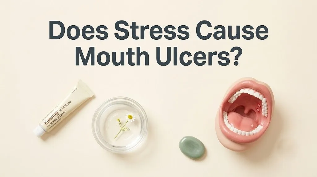
The first-line treatment is a topical corticosteroid. A dentist or physician can prescribe triamcinolone acetonide 0.1% dental paste (Kenalog in Orabase). A small amount is dabbed directly onto the dry ulcer with a clean finger or cotton swab before bed. The steroid suppresses the local TNF-alpha-driven inflammation and reduces the pain and redness. The paste adheres to the ulcer and provides a physical barrier against further irritation. The key is to apply it at the earliest sign of the ulcer, the prodromal burning or tingling phase, before the crater fully forms.
Over-the-counter options include amlexanox 5% oral paste (Aphthasol), which is the only FDA-approved topical treatment specifically for aphthous ulcers. It accelerates healing time by approximately one day. Benzydamine hydrochloride 0.15% mouthwash (Tantum Verde) provides pain relief by acting as a local anesthetic and anti-inflammatory. A warm salt water rinse, a half teaspoon of salt in a cup of warm water, swished for 30 seconds three to four times a day, provides a gentle, antimicrobial cleansing that is better than using a commercial mouthwash, which often contains alcohol and can sting and irritate the ulcer.
Do not apply silver nitrate sticks or other chemical cautery agents sold as “canker sore cures” without professional guidance. These chemically burn the ulcer, destroying the nerve endings and providing temporary pain relief, but they also destroy the healthy tissue at the edges of the ulcer, which can delay healing. A dentist can properly debride and treat a severe ulcer. Self-cauterization is risky.
Key Takeaway: A prescription steroid paste dabbed on the ulcer at the first tingle is the single most effective treatment. The over-the-counter alternatives are salt water rinses and amlexanox paste.
best products for stress mouth ulcer relief
The best products for stress mouth ulcer relief are categorized into pain-relieving barriers, anti-inflammatory agents, and antimicrobial rinses, with the specific product choice depending on the location and severity of the ulcer. The goal is to create a protective film over the exposed nerve endings to eliminate the constant, stinging pain that makes eating, drinking, and speaking miserable.
Barrier products form a physical gel or patch over the ulcer. Zilactin-B is a gel that forms a bioadhesive film. Canker Cover is a dissolvable patch that adheres to the ulcer and provides up to 12 hours of coverage. These products do not heal the ulcer faster, but they provide significant pain relief by sealing the nerve endings from air, saliva, and food. They are best for ulcers on the inner lip or cheek where they can adhere without being washed away by the tongue.
Antimicrobial and anti-inflammatory mouthwashes are the second category. A chlorhexidine gluconate 0.12% mouthwash, used twice daily, reduces the bacterial load on the ulcer and prevents secondary infection. It is prescription-only in the US and should not be used for more than two weeks due to the risk of tooth staining. Benzydamine hydrochloride mouthwash is an over-the-counter anti-inflammatory and local anesthetic. It reduces pain within 15 minutes of rinsing.
For ulcers on the tongue, a topical spray is often easier to apply than a paste. Difflam spray delivers a metered dose of benzydamine directly to the tongue ulcer. For ulcers on the soft palate or near the throat, a gargle with soluble aspirin or benzydamine provides relief. The product must be able to reach the ulcer. A paste that works perfectly on the lip will be useless for an ulcer at the back of the tongue.
Avoid products that contain alcohol, as they sting intensely and dry out the mucosa, prolonging healing. Avoid sodium lauryl sulfate (SLS) toothpaste, which strips the protective mucin layer. Biotene and Sensodyne Pronamel are SLS-free options.
how to prevent stress mouth ulcers
To prevent stress mouth ulcers, you must simultaneously address the psychological driver by reducing the frequency and intensity of the HPA axis activation and address the local oral environment by switching to an SLS-free toothpaste, identifying and avoiding dietary triggers, and correcting any underlying nutritional deficiencies. Prevention is a two-front war: the systemic stress response and the local oral environment.
The systemic strategy is stress management. The stress that causes mouth ulcers is typically chronic or repeated acute stress, the exam period, the work deadline, the relationship conflict. Evidence-based stress reduction techniques that lower cortisol and increase parasympathetic tone are the preventive intervention. A 2023 randomized controlled trial published in the Journal of Behavioral Medicine found that an 8-week mindfulness-based stress reduction program reduced the incidence of RAS by over 50 percent in chronic sufferers compared to a waitlist control.
The local strategy is to harden the oral mucosa against the immune suppression you cannot completely eliminate. Switch to an SLS-free toothpaste. Sodium lauryl sulfate is a detergent that strips the protective salivary mucin layer and can directly irritate the oral epithelium. For a person prone to stress ulcers, this is an unnecessary chemical injury. Keep a food diary to identify dietary triggers. The most common offenders are chocolate, nuts, coffee, strawberries, and highly acidic foods like tomatoes and citrus.
Correct nutritional deficiencies with targeted supplementation, not a multivitamin. Have your B12, ferritin, and folate levels checked. If any are low, replace them with a specific supplement. A 2022 study in Oral Diseases found that B12 supplementation alone reduced RAS episodes by over 60 percent in deficient patients. The supplement is treating the co-factor, not the stress, but the ulcer does not know the difference.
can stress cause ulcers in the mouth repeatedly
Stress can cause ulcers in the mouth repeatedly, and this is the hallmark of recurrent aphthous stomatitis, or RAS, where the patient experiences predictable, cyclical outbreaks of ulcers that correlate with periods of heightened psychological stress, menstrual cycles, or both. If you find yourself reaching for the same tube of steroid paste every time a big project looms at work, you have a stress-triggered RAS pattern.
The mechanism of recurrence is not that each new stressor creates new ulcers from scratch. It is that the baseline allostatic load, the cumulative physiological burden of chronic stress, keeps the oral mucosal immune system in a constantly suppressed state. The sIgA is chronically low. The Th1 response is blunted. The epithelium is thin. The mouth is a minefield of potential ulcer sites, just waiting for a trigger. A single stressful event does not cause the recurrence. The constant background stress creates the vulnerability. The acute stressor tips the scale.
Patients with RAS often have a predictable latency period. The ulcers appear not during the absolute peak of the stressor, but 24 to 72 hours after, during the “let-down” period. This is because the acute cortisol surge suppresses the initial inflammatory response that would create the ulcer. The immune system is suppressed. When the cortisol spike drops, the immune system rebounds, and the inflammatory response to the micro-wound that occurred during the stress suddenly erupts. The ulcer appears on the first day of vacation, a classic clinical observation.
Management of recurrent stress ulcers requires a long-term strategy. A prophylactic SLS-free toothpaste, consistent B12 status, daily stress management, and a prescription steroid paste on hand to apply at the very first prodromal tingle. The goal is not to eliminate every ulcer forever. That may not be possible if you have the genetic predisposition. The goal is to reduce the frequency from monthly to annually.
Key Takeaway: If your mouth ulcers appear like clockwork after every stressful period, you have recurrent aphthous stomatitis, and you need a long-term prevention plan.
when to see a dentist or doctor for mouth ulcers
You should see a dentist for mouth ulcers when a single ulcer persists for more than two weeks without healing, when you develop more than five ulcers at once, when the ulcers are larger than one centimeter in diameter, or when the ulcers are accompanied by genital ulcers, skin lesions, joint pain, fever, or gastrointestinal symptoms. These features suggest a systemic disease process, not simple stress-induced aphthous stomatitis.
A non-healing ulcer is the most critical red flag. The oral mucosa heals rapidly. Any ulcer that has not shown clear signs of healing within 14 days requires a biopsy to rule out oral squamous cell carcinoma. This is not a rare precaution. Delayed diagnosis of oral cancer is a significant cause of mortality because the early lesion is painless and ignored. A stress ulcer heals. A cancer ulcer does not.
The systemic disease red flags include complex aphthosis, where the patient has large, deep, scarring ulcers inside the mouth and on the genitals. This is a hallmark of Behcet’s disease, a systemic vasculitis that requires management by a rheumatologist. Crohn’s disease can present with oral ulcers that precede any gastrointestinal symptoms. A patient with recurrent mouth ulcers and unexplained abdominal pain, diarrhea, or weight loss needs a referral to a gastroenterologist.
The initial evaluation can be done by your primary care dentist. They will take a thorough history, examine the lesion, and palpate the neck for lymph nodes. If the dentist is concerned, they will refer you to an oral medicine specialist, a dentist with advanced training in the diseases of the oral mucosa. The specialist can perform a biopsy in the chair, order the relevant blood tests, and coordinate care with a rheumatologist or gastroenterologist if indicated. Do not let a non-healing mouth ulcer go unevaluated.
Frequently Asked Questions About Stress and Mouth Ulcers
Why do I get mouth ulcers when I’m stressed?
You get mouth ulcers when stressed because cortisol and epinephrine suppress your oral immune defense, specifically depleting salivary IgA and shifting your T-helper cell balance away from the type that protects your mouth lining.
This immune suppression allows the normal micro-trauma from eating or accidentally biting your cheek to develop into a full ulcer instead of healing quietly.
The ulcer appears 24 to 72 hours after the stress peak during the “let-down” phase.
What does a stress ulcer look like in the mouth?
A stress mouth ulcer looks like a small, round, or oval crater with a flat, white-yellow base and an intensely red, inflamed border on the movable tissue inside your lips, cheeks, or under your tongue.
It is typically less than one centimeter across and is exquisitely painful, with the pain often disproportionate to the small size of the sore.
It does not blister, and it does not crust unless it is on the dry surface of the lip.
How long do stress mouth ulcers last?
A stress mouth ulcer typically lasts 7 to 14 days, with the most intense pain occurring in the first 3 to 5 days, after which the white-yellow base is gradually replaced by healthy pink tissue.
If an ulcer persists beyond two weeks without any sign of healing, it requires evaluation by a dentist or oral medicine specialist.
The healing time can be shortened by applying a topical corticosteroid paste at the earliest tingling sensation.
Can stress cause ulcers on the tongue?
Yes, stress can cause ulcers on the lateral borders and tip of the tongue through both the immune suppression mechanism and through the mechanical trauma of unconscious tongue biting during concentration or sleep.
The tongue is also prone to a stress-related condition called geographic tongue, where smooth, red, map-like patches appear and can be sore.
A non-healing ulcer on the side of the tongue needs prompt dental evaluation to rule out oral cancer, especially in smokers.
What is the fastest way to heal a stress mouth ulcer?
The fastest way to heal a stress mouth ulcer is to apply a prescription topical corticosteroid paste, such as triamcinolone acetonide 0.1%, directly to the ulcer at the very first prodromal burning or tingling sensation.
This suppresses the TNF-alpha-driven inflammation before the crater fully forms and can shorten healing time by several days.
A barrier patch and warm salt water rinses can protect the ulcer and reduce bacterial irritation while it heals.
How do I know if my mouth ulcer is from stress or something serious?
You know a mouth ulcer is likely from stress if it is small, heals within two weeks, occurs on the movable tissue inside your mouth, and appears in a clear temporal relationship with a stressful period.
It may be something more serious if it persists beyond two weeks without healing, is larger than one centimeter, is accompanied by genital sores, skin lesions, joint pain, or abdominal symptoms, or if you have never had ulcers before and suddenly develop them after age 40.
A dentist or oral medicine specialist can differentiate a stress ulcer from oral cancer, autoimmune disease, or a viral infection.
The mouth ulcer you get after a terrible week is not a random inconvenience. It is a visible, painful manifestation of your HPA axis altering the immune defense of your oral cavity at the cellular level. Your body is not betraying you. It is reporting to you, with a precise, localized biological signal, that the stress you are under has crossed a threshold where it is now suppressing your mucosal immunity.
The single most practical step you can take tonight is to apply a protective barrier paste or patch to the ulcer so you can eat and sleep without pain. Tomorrow, make the switch to an SLS-free toothpaste. This one change can independently reduce your ulcer frequency. If you are getting these ulcers repeatedly, ask your primary care provider for a blood draw to check your B12, ferritin, and folate. Correcting a deficiency is a high-impact, evidence-based intervention.
Your stress is real. The ulcer is real. The connection between them is scientifically mapped, from the brain’s perception of a threat down to the T-cell signaling failure in your cheek. You now have the knowledge and the specific tools to break that chain at both ends. Your mouth deserves to be a comfortable, pain-free environment, even when your life is chaotic.