Stress Test vs Stress Echo: 2026 Key Differences Guide
The fundamental difference between a standard stress test and a stress echo is that a stress test monitors the heart’s electrical activity with an electrocardiogram during exercise to detect signs of ischemia, while a stress echo adds an ultrasound imaging component, an echocardiogram, performed immediately before and after the exercise, which visualizes the heart muscle’s squeezing motion to detect areas of the wall that are not moving properly due to a lack of blood flow. The stress test listens to the heart’s electrical rhythm. The stress echo watches the heart’s mechanical dance.
A 2023 meta-analysis published in the Journal of the American College of Cardiology found that a stress echocardiogram has a sensitivity of approximately 80 to 85 percent for detecting a flow-limiting coronary blockage, compared to a sensitivity of 65 to 70 percent for a standard exercise EKG stress test. The stress echo detects the wall motion abnormality, which occurs earlier in the ischemic cascade than the electrical changes seen on the EKG.
This guide will explain the exact mechanistic difference between the two tests, map out the ischemic cascade that makes the stress echo more accurate, and provide a clear decision framework for which test is appropriate for your specific clinical situation. If you have been told you need a “stress test” and are unsure which type you are getting, this is your definitive comparison.
If you are in crisis or experiencing thoughts of self-harm, contact the 988 Suicide and Crisis Lifeline by calling or texting 988 at any time. This service is free, confidential, and available 24 hours a day.
what is the difference between stress test and stress echo
The difference between a stress test and a stress echo is the technology used to detect the heart’s response to stress. A stress test uses a continuous 12-lead electrocardiogram, or EKG, to monitor the electrical activity of the heart for characteristic ST segment changes that indicate ischemia. A stress echo uses an ultrasound probe placed on the chest to capture live video images of the heart muscle contracting, looking for a wall motion abnormality.
The EKG-based stress test is like listening to the engine of a car for a misfire. The stress echo is like opening the hood and watching the pistons move. Both can detect a problem, but the visual inspection is often more sensitive.
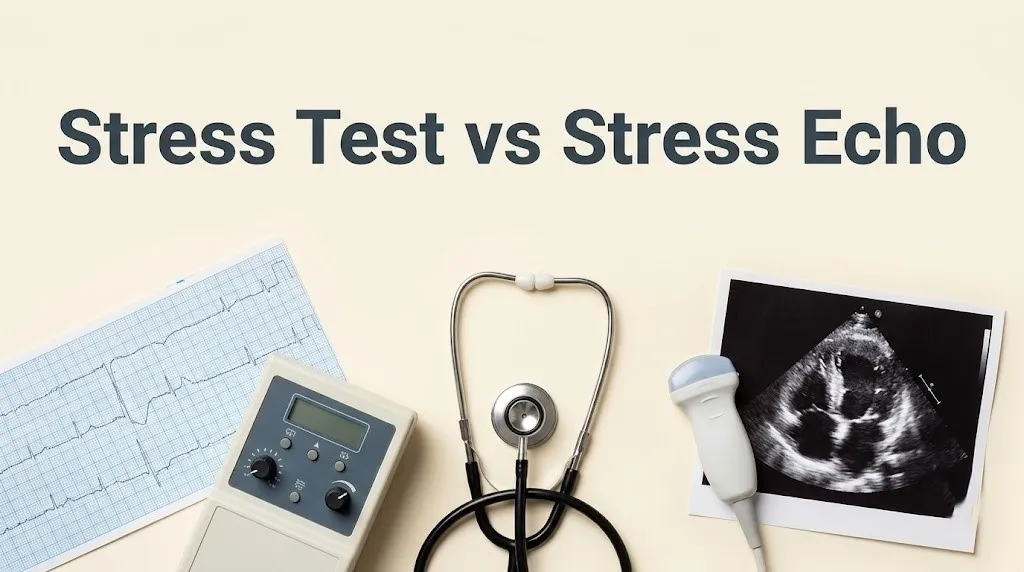
The standard stress test is the older, simpler, and more widely available technology. It requires a treadmill, an EKG machine, and a trained technician. It is the appropriate first-line screening test for a patient with a normal baseline EKG who can exercise adequately and has a low-to-intermediate pre-test probability of disease.
The stress echo adds an echocardiogram, which is performed by a cardiac sonographer. The sonographer takes images of the heart at rest, and then immediately after the patient steps off the treadmill, the sonographer takes another set of images while the heart is still beating rapidly from the exertion. The two sets of images are compared side-by-side.
Key Takeaway: A stress test records your heart’s electrical rhythm during exercise. A stress echo records a movie of your heart muscle squeezing at rest and immediately after exercise.
stress test ekg vs stress echo ultrasound
The stress test EKG relies on the detection of ST segment depression on the electrocardiogram. The stress echo ultrasound relies on the visualization of a regional wall motion abnormality on the echocardiogram. The two modalities detect ischemia at different points in the ischemic cascade.
The EKG measures the voltage difference between electrodes placed on the skin. When a region of the heart muscle becomes ischemic, the electrical repolarization of the cells is altered. This produces a characteristic depression of the ST segment on the EKG tracing. The ST segment must be depressed by at least 1 millimeter in multiple contiguous leads to be considered a positive test.
The echocardiogram uses high-frequency sound waves to create a real-time image of the heart. The sonographer views the four chambers of the heart, the valves, and the motion of the left ventricular walls. A normal heart wall thickens and moves inward during contraction. An ischemic heart wall becomes hypokinetic, moving less vigorously, or akinetic, not moving at all.
The wall motion abnormality is a direct, visible consequence of the lack of oxygen delivery to that segment of the muscle. It is a mechanical failure that precedes the electrical change on the EKG.
how does a stress echo detect ischemia
A stress echo detects ischemia by capturing ultrasound images of the left ventricle at rest and immediately after peak exercise, and then comparing the motion of each segment of the heart wall. A segment that contracts normally at rest but becomes sluggish or motionless immediately after exercise is a positive test for a flow-limiting blockage in the coronary artery that supplies that segment.
The left ventricle is divided into 17 standardized segments. Each segment is supplied by one of the three major coronary arteries: the left anterior descending, the circumflex, or the right coronary artery. The cardiologist can map the wall motion abnormality to the specific diseased artery.
The ischemic cascade explains the physiology. When blood flow to a region of the heart muscle is reduced, the first abnormality is a perfusion defect, seen on a nuclear scan. The second is a diastolic dysfunction, a stiffness of the relaxing heart muscle. The third is the wall motion abnormality, which the stress echo detects. The fourth is the ST segment depression on the EKG. The fifth is chest pain.
Because the stress echo detects the wall motion abnormality earlier in the cascade than the EKG detects the ST change, the stress echo is more sensitive. It can detect a milder degree of ischemia.
standard stress test sensitivity and accuracy
A standard exercise treadmill stress test, without imaging, has a sensitivity of approximately 65 to 70 percent and a specificity of approximately 70 to 75 percent for detecting a flow-limiting coronary artery blockage. The accuracy is highly dependent on the patient’s baseline EKG and their ability to achieve an adequate heart rate.
The sensitivity is limited by the fact that some patients never develop the diagnostic ST segment depression, even with significant ischemia. The false-negative rate is approximately 30 percent.
The specificity is limited by the fact that many conditions other than coronary artery disease can cause ST segment depression, including left ventricular hypertrophy, a bundle branch block, and even anxiety and hyperventilation. The false-positive rate is also approximately 30 percent.
A 2022 study in Circulation found that the diagnostic accuracy of the standard stress test was significantly lower in women than in men, primarily due to a higher rate of false-positive EKG changes in women.
stress echo advantages over treadmill test
The stress echo has several specific advantages over the standard treadmill test. It has a higher sensitivity and specificity. It can localize the ischemic territory to a specific coronary artery. It can evaluate the heart valves and the overall ejection fraction. It is not affected by baseline EKG abnormalities.
The improved accuracy is the primary advantage. A stress echo is approximately 15 to 20 percentage points more sensitive than a standard EKG test. This means it misses fewer significant blockages.
The ability to localize the ischemia is a major advantage for planning further treatment. If the stress echo shows an anterior wall motion abnormality, the cardiologist knows the left anterior descending artery is likely the culprit.
The stress echo also provides information about the heart valves, particularly the aortic and mitral valves, and the global systolic function, the ejection fraction. This information is not available from the EKG alone.
the ischemic cascade and stress echo
The ischemic cascade is the sequence of pathophysiological events that occur when the heart muscle is deprived of oxygen. Understanding this cascade is the key to understanding why a stress echo is more sensitive than a standard stress test. The wall motion abnormality occurs before the ST segment change.
The cascade begins with a reduction in coronary blood flow. The myocardial perfusion becomes heterogeneous. This perfusion defect can be detected by a nuclear scan.
Next, the heart muscle’s ability to relax during diastole is impaired. This diastolic dysfunction is subtle and not clinically detectable on a standard test.
Next, the heart muscle’s ability to contract during systole is impaired. This is the regional wall motion abnormality. The stress echo detects the wall motion abnormality, which typically occurs within seconds of the onset of ischemia.
Only after the wall motion abnormality is established does the EKG begin to show the ST segment depression. The electrical change is a later phenomenon. The stress echo is therefore detecting an earlier, more sensitive marker of ischemia.
Key Takeaway: The stress echo sees the heart muscle failing to squeeze, which happens first. The stress test sees the electrical signal changing, which happens later.
stress echo vs nuclear stress test
A stress echo uses ultrasound to image the heart, involves no ionizing radiation, and has a sensitivity of 80 to 85 percent. A nuclear stress test uses a radioactive tracer to image the heart’s perfusion, involves a radiation dose of 8 to 15 mSv, and has a sensitivity of 85 to 90 percent.
The nuclear stress test is slightly more sensitive than the stress echo, particularly for detecting mild, single-vessel disease. The perfusion defect is the earliest event in the ischemic cascade.
However, the nuclear stress test involves a significant radiation dose, which is a consideration for younger patients, particularly women. It is also more expensive and takes longer to perform.
The stress echo is often the preferred initial imaging test because it avoids radiation, provides structural information about the valves and the pericardium, and is widely available.
which is better stress test or stress echo
A stress echo is generally better than a standard stress test for most patients because of its higher sensitivity and specificity, its ability to localize the ischemia, and its ability to provide additional structural information about the heart. However, a standard stress test is a reasonable, lower-cost screening test for a patient with a low pre-test probability of disease and a normal baseline EKG.
The American College of Cardiology guidelines recommend an imaging stress test, either a stress echo or a nuclear test, for patients with an abnormal baseline EKG, patients who have had a prior inconclusive standard stress test, and patients with a higher pre-test probability of disease.
The choice between a stress echo and a standard stress test is a decision that should be made by the cardiologist based on the individual patient’s clinical profile.
stress echo for women and difficult patients
A stress echo is particularly advantageous for women and for patients with baseline EKG abnormalities, obesity, or lung disease. The standard stress test has a notoriously high false-positive rate in these populations.
Women are more likely to have a false-positive ST segment depression on a standard stress test. The exact mechanism is not fully understood, but it may be related to estrogen and the smaller size of the coronary arteries.
Patients with a left bundle branch block, left ventricular hypertrophy, or a paced rhythm have baseline EKG abnormalities that make the ST segment uninterpretable. A stress echo is essential for these patients.
Obese patients and patients with chronic obstructive pulmonary disease often have poor acoustic windows for a resting echocardiogram, but the stress echo can still be performed with contrast agents that improve the definition of the endocardial border.
stress test false positive vs stress echo
A false-positive stress test occurs when the EKG shows ST segment depression, but the patient does not have a significant coronary blockage on the angiogram. The stress echo has a lower false-positive rate because the wall motion abnormality is a more specific marker of ischemia than the ST segment change.
A false-positive standard stress test can lead to unnecessary anxiety, additional testing, and even an unnecessary invasive angiogram. The stress echo reduces the probability of this pathway.
The false-positive rate of a standard stress test is approximately 25 to 30 percent. The false-positive rate of a stress echo is approximately 10 to 15 percent.
If a patient has had a false-positive stress test in the past, a stress echo is the appropriate next test.
what happens during a stress echo procedure
During a stress echo, the patient first undergoes a resting echocardiogram. The sonographer places the ultrasound probe on the chest and captures images of the heart from multiple views. The patient then exercises on a treadmill, with the speed and incline increasing every 3 minutes.
At peak exercise, the patient quickly steps off the treadmill and lies back down on the exam table. The sonographer immediately re-acquires the images while the heart is still beating rapidly. The entire process takes about 45 to 60 minutes.
If the patient is unable to exercise, a pharmacological stress agent, such as dobutamine, is infused through an IV. The dobutamine increases the heart rate and the contractility, mimicking the effect of exercise.
cost and availability of stress test vs echo
A standard stress test is less expensive and more widely available than a stress echo. It can be performed in a primary care clinic or a community hospital. A stress echo requires a cardiac sonographer and a cardiologist to interpret the images, and it is typically performed in a cardiology clinic or a hospital.
The cost difference is several hundred dollars. The standard stress test is the most cost-effective initial screening test for a low-risk population.
Insurance coverage for both tests is generally good when the test is ordered for an appropriate clinical indication.
preparing for a stress test or stress echo
The preparation for a stress test and a stress echo is identical. The patient must fast for 4 to 6 hours before the test. Caffeine must be avoided for 12 to 24 hours. Specific cardiac medications, such as beta-blockers, may need to be held for 24 to 48 hours.
The patient should wear comfortable clothing and walking shoes for the treadmill. Do not apply lotion or powder to the chest.
stress echo vs exercise stress test results
The results of a stress echo are reported as the presence or absence of an inducible wall motion abnormality, the location and severity of the abnormality, and the global ejection fraction. The results of a standard stress test are reported as the presence or absence of significant ST segment depression, the heart rate and blood pressure response, and the exercise capacity.
An abnormal stress echo is a more definitive finding than an abnormal standard stress test. It is more likely to lead to a recommendation for an invasive angiogram.
when is a stress echo medically necessary
A stress echo is medically necessary when the patient has an abnormal baseline EKG that would make the standard stress test uninterpretable, when the patient has had a prior inconclusive or false-positive standard stress test, or when the patient has a high pre-test probability of coronary artery disease.
A stress echo is also indicated when the cardiologist needs additional information about the heart valves or the ejection fraction.
can a stress echo replace an angiogram
A stress echo cannot replace a coronary angiogram. A stress echo is a non-invasive functional test that detects the downstream effect of a blockage on the heart muscle. An angiogram is an invasive anatomical test that directly visualizes the blockage inside the coronary artery.
A normal stress echo is highly reassuring and often means an angiogram is unnecessary. An abnormal stress echo is a strong indication for proceeding to an angiogram.
Key Takeaway: The stress echo is the gatekeeper. It tells you if a blockage is likely causing a problem. The angiogram is the inside look that can fix the blockage.
Frequently Asked Questions About Stress Tests and Stress Echos
Is a stress echo better than a regular stress test?
Yes, a stress echo is more accurate than a regular stress test.
It has a sensitivity of 80 to 85 percent, compared to 65 to 70 percent for a standard EKG test.
The stress echo detects a wall motion abnormality, which occurs earlier in the ischemic cascade than the EKG changes.
Why would a doctor order a stress echo instead of a stress test?
A doctor would order a stress echo if the patient has an abnormal baseline EKG, is a woman, or has a higher pre-test probability of coronary artery disease.
The stress echo is less likely to produce a false-positive result.
It also provides additional information about the heart valves and the pumping function.
Can a stress echo detect blockages better?
A stress echo can detect the physiological consequence of a flow-limiting blockage more accurately than a standard stress test.
It detects fewer false negatives, meaning it is better at ruling out significant disease.
It cannot directly visualize the plaque like an angiogram.
Does a stress echo have more risks?
A stress echo has the same low risk profile as a standard stress test.
The exercise carries a very small risk of triggering a heart rhythm abnormality or a heart attack.
The ultrasound itself is completely safe and involves no radiation.
Is a stress echo more expensive?
A stress echo is more expensive than a standard stress test because it requires a cardiac sonographer and specialized ultrasound equipment.
The cost difference is several hundred dollars.
Insurance generally covers both tests when medically indicated.
What is the difference between a nuclear stress test and a stress echo?
A nuclear stress test uses a radioactive tracer to map blood flow and has a sensitivity of 85 to 90 percent.
A stress echo uses ultrasound to watch the heart muscle squeeze and has a sensitivity of 80 to 85 percent.
The nuclear test has a slightly higher sensitivity but involves a radiation dose of 8 to 15 mSv.
The choice between a stress test and a stress echo is a choice between listening to the heart’s electrical rhythm and watching the heart’s mechanical dance. The EKG listens for the ST segment depression. The ultrasound watches for the wall motion abnormality. The wall motion fails first. The electrical signal changes later. The stress echo is therefore the more sensitive test, particularly for women and for patients with tricky baseline EKGs.
If your cardiologist has ordered a stress echo, you now understand why. The test is a higher-resolution look at your heart’s function under stress. It avoids radiation, it is widely available, and it provides a wealth of structural information beyond just the ischemia.
Prepare by fasting and avoiding caffeine. Show up ready to walk on the treadmill. The sonographer will take the pictures at rest and immediately after you step off. The images will be compared. The wall motion will be graded. You will walk out with a clear answer about whether your heart muscle is getting the blood it needs when it works hard.