How to Tell If You Have a Stress Fracture: 2026 Guide
To tell if you have a stress fracture, you need to identify a very specific pattern of pain: a sharp, focal, pinpoint tenderness directly on a bone that worsens with impact activity, improves transiently with rest, and hurts at night or with the vibration of a tuning fork if applied. The classic self-assessment is the hop test. If you can hop pain-free on the affected leg, a stress fracture is unlikely. If hopping produces a sharp, deep pain at a specific bony point, a stress fracture is highly probable.
A stress fracture is not a crack from a single traumatic fall. It is the endpoint of a biological failure. Your bone is living tissue that constantly remodels itself, breaking down old bone and laying down new bone. When you increase your training load too quickly, the osteoclasts resorb bone faster than the osteoblasts can rebuild it. The bone weakens. Microcracks form. This is a stress reaction, the precursor. If the loading continues, the microcracks coalesce into a frank cortical fracture.
This guide will teach you exactly how to self-assess your injury, grade its severity, and understand why the “stress” in stress fracture is not just mechanical but also deeply neuroendocrine. Chronic psychological stress and the associated elevation of cortisol directly weakens your bone microarchitecture. We will cover the specific pain patterns, the critical MRI grading system, and the exact moment you need to see an orthopedic surgeon.
how to tell if you have a stress fracture
You can tell if you have a stress fracture by the presence of a focal, “one-finger” point of exquisite tenderness directly on a bone, pain that worsens with running or jumping and improves within minutes of stopping, and a positive response to the hop test, which produces sharp, deep pain at the suspected fracture site. Unlike a muscle strain, which hurts when you stretch or squeeze the muscle belly, a stress fracture hurts specifically when you press on the hard, superficial surface of the bone itself.
The pain progression is predictable. In the earliest stage, a stress reaction, the pain occurs only after activity, typically at the end of a run or training session. The athlete often dismisses it as a tight muscle. As the stress reaction deepens into a developing fracture, the pain is present at the start of activity, “warms up” and feels better during the middle, and then returns intensely after the activity stops. This is the “warm-up phenomenon.” When the fracture is complete, the pain is constant, present at rest, and often wakes the patient from sleep. Any pain at night is an urgent warning sign.
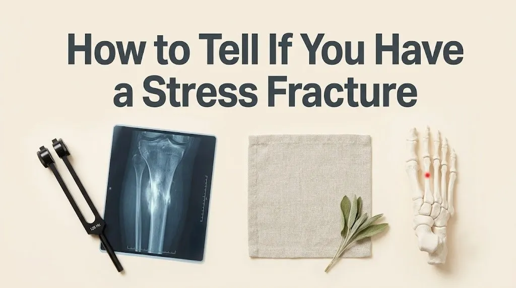
The location of the tenderness is diagnostic. Common sites include the second or third metatarsal shaft in the foot, the distal tibia on the inner shin, the navicular bone on the top of the arch, the femoral neck in the groin, and the pars interarticularis in the low back. You can typically cover the pain of a stress fracture with the pad of a single fingertip. A muscle strain or shin splints (medial tibial stress syndrome) produces a diffuse, achy tenderness over a longer segment of the soft tissue.
You can perform a rough tuning fork test at home. Strike a 128 Hz tuning fork against a hard surface and press the base against the suspected bone. The high-frequency vibration travels through the bone and vibrates the edges of the fracture crack, producing a sharp, lancinating pain. A positive tuning fork test is highly specific for a cortical fracture. It does not hurt a strain or tendonitis. If you have pinpoint bony tenderness, a positive hop test, and a positive tuning fork test, a stress fracture is the working diagnosis until an MRI proves otherwise.
Key Takeaway: A stress fracture hurts on the bone, not the muscle. Press firmly with one finger. If it makes you wince, you need an X-ray and likely an MRI.
do i have a stress fracture
You likely have a stress fracture if you have recently increased your training volume, intensity, or surface hardness, and you now have a specific, focal bone pain that has progressed through the predictable stages from “pain after activity” to “pain during activity” to “pain at rest.” The quickest way to answer the question is to stop running and start walking. If walking on a hard surface produces the same sharp, deep pain at the same bony point, a stress fracture is very likely.
The history is as important as the physical exam. Stress fractures are the classic “too much, too soon” injury. Did you double your weekly mileage in two weeks? Did you switch from treadmill running to concrete pavement? Did you start a jump-heavy plyometric program? Did you join a military boot camp? The bone requires approximately 6 to 8 weeks to adapt to a new mechanical load. A 2023 study in the American Journal of Sports Medicine found that runners who increased their mileage by more than 30 percent in a single week had a 4-fold higher risk of developing a bone stress injury.
Your body type and lifestyle also provide clues. Are you a female with irregular or absent menstrual periods? The loss of estrogen removes a primary protective hormone for bone, leading to accelerated resorption. Are you under-fueling? A calorie deficit, particularly a low-carbohydrate intake, suppresses insulin-like growth factor 1, a key bone-building hormone. Are you a smoker? Nicotine is a direct vasoconstrictor that reduces blood flow to the bones by up to 40 percent.
The “pinch test” is a self-exam tool. Gently squeeze the injured bone from both sides. For a metatarsal, squeeze the forefoot. For the calcaneus, squeeze the heel from both sides. If the compressive pressure produces a deep, nauseating pain, it is a positive squeeze test and highly suggestive of a stress fracture. Normal muscles and tendons do not produce this compressive bone pain.
how do i know if i have a stress fracture
You know if you have a stress fracture definitively through imaging, specifically an MRI, which can detect a bone stress injury within 24 to 48 hours of symptom onset, long before an X-ray shows the characteristic “dreaded black line” of a frank fracture. The clinical tests, the history, the hop test, and the tuning fork test give you a high probability. The MRI gives you the diagnosis.
The diagnostic path typically begins with an X-ray. However, it is critical to understand that an X-ray is negative in up to 70 percent of early stress fractures. The microcracks and bone marrow edema are not visible on plain radiographs for 2 to 4 weeks, when the healing response produces a visible periosteal reaction or a sclerotic line at the fracture site. A normal X-ray does not rule out a stress fracture. A 2024 systematic review in the American Journal of Sports Medicine confirmed that MRI is the gold standard with a sensitivity and specificity approaching 100 percent.
The MRI sequence is specific. The technologist will perform a T1-weighted and a STIR (Short Tau Inversion Recovery) sequence. The STIR sequence suppresses the signal from fat and makes fluid, such as the edema in a stressed bone, glow bright white. The orthopedic surgeon will grade the injury on the Fredericson MRI grading system, which ranges from Grade 1 (mild periosteal edema) to Grade 4 (a clear cortical fracture line with severe bone marrow edema).
You do not need to interpret the MRI yourself. The radiologist’s report will specify the grade. A Grade 1 or 2 injury is a stress reaction. It is treated non-operatively with rest. A Grade 3 or 4 injury is a stress fracture. A high-grade injury in a high-risk site, such as the femoral neck or the navicular, may require surgical fixation with a screw.
Key Takeaway: An MRI is the only definitive way to know. An X-ray will be falsely negative in the first few weeks. Insist on an MRI if the pain persists.
what does a stress fracture feel like
A stress fracture feels like a deep, dull, aching pain localized to a specific bone that is exacerbated by impact and relieved by offloading. It is often described as a “toothache in the bone.” The bone aches constantly at rest in the later stages. The pain is not sharp like a knife cut, but a deep, nauseating, gnawing ache that is distinctly different from the burning or stretching sensation of a tendon or muscle injury.
The pain has a mechanical rhythm. Walking across a hard floor, stepping off a curb, or pushing off the foot during a stride generates a wave of deep pain. Resting, elevating the leg, and applying ice provides temporary relief. The pain is frequently worse at night. During sleep, the sympathetic nervous system tone drops, and the pro-inflammatory cytokines that have accumulated in the damaged bone tissue stimulate the nerve endings without the distraction of movement. This nocturnal bone pain is a critical clinical sign.
The pain is highly localized. A patient with a tibial stress fracture can point to the exact spot on the shin with a single finger. A patient with shin splints will rub a palm over a large, diffuse area of the inner shin. A patient with a femoral neck stress fracture will feel the pain deep in the groin or the front of the hip, and it may radiate to the knee. A metatarsal stress fracture feels like a stone bruise that never goes away, a painful lump under the ball of the foot.
The pain does not respond to typical soft-tissue treatments. Foam rolling, massage, and stretching do not help a stress fracture. In fact, massage directly over the fracture site can be agonizing. This is a key differentiator from a muscle strain.
stress reaction vs stress fracture symptoms
A stress reaction is the earliest phase of a bone stress injury, presenting with pain only after activity, no pain at rest, and a negative X-ray, while a stress fracture is a later stage with pain during activity, pain at rest, and a visible fracture line on MRI. The symptoms exist on a spectrum of severity.
The stress reaction phase is characterized by bone marrow edema without a cortical break. The bone is bruised and swollen on the inside. The pain is typically a dull ache that appears several hours after the activity and is gone by the next morning. The athlete can often “run through” a stress reaction, although this is exactly what converts it into a true fracture. The clinical exam reveals some point tenderness, but the hop test may be equivocal or only mildly positive.
The stress fracture phase is characterized by a cortical crack. The pain is now present at the start of the activity, may warm up slightly, but never fully disappears. It is present during walking and can be sharp with any mis-step. The hop test is severely positive. The tuning fork test is positive. The X-ray may still be negative, but the MRI will show a Grade 3 or 4 injury with a visible fracture line through the cortex.
The treatment differs drastically between the two. A stress reaction (Grade 1-2) can often be managed with a period of relative rest and activity modification for 4 to 6 weeks, followed by a gradual return to running. A stress fracture (Grade 3-4) requires a period of strict non-weight-bearing or bracing for 6 to 8 weeks. A stress reaction can be managed by a physical therapist. A stress fracture must be managed by an orthopedic surgeon.
| Feature | Stress Reaction | Stress Fracture |
|---|---|---|
| Pain Onset | After activity only | During activity, at rest, at night |
| Hop Test | Negative or mild | Sharply positive |
| X-ray | Normal | May be normal, or show callus/line |
| MRI Grading | Grade 1-2 (edema only) | Grade 3-4 (cortical break) |
| Treatment | Relative rest, 4-6 weeks | Non-weight-bearing, 6-8 weeks |
| Provider | Physical Therapist | Orthopedic Surgeon |
what does a femur stress fracture feel like
A femur stress fracture feels like a deep, achy, sickening pain in the groin, the front of the hip, or the deep buttock that is exquisitely worsened by pivoting, single-leg stance, or raising the leg against resistance, and it often refers pain to the knee. The femoral neck is a high-risk site, and a missed femoral neck stress fracture can be a catastrophic, career-ending injury.
The pain is insidious. It starts as a vague groin ache after a long run or a heavy squat session. The athlete may stretch the groin, thinking it is a tight adductor muscle. The pain progresses. It becomes uncomfortable to lie on the affected side at night. Rolling over in bed produces a deep twinge. Walking up stairs or an incline is painful.
The specific clinical test for a femoral neck stress fracture is the log roll test. Lie on your back with your leg straight. Have someone else gently roll your leg inward and outward from the ankle. If the rotational movement produces a deep, sharp pain in the groin, the hip joint or the femoral neck is the source.
This fracture is dangerous because it can displace. A tension-side stress fracture, on the superior aspect of the femoral neck, is under tensile load and is at high risk of displacing, cutting off the blood supply to the femoral head, and causing avascular necrosis. This requires emergent surgical fixation with screws. A compression-side fracture, on the inferior aspect, is usually stable and can be treated non-operatively.
A displaced femoral neck stress fracture is a surgical emergency. The pain is sudden, severe, and the patient cannot bear weight. If you have deep groin pain and a positive log roll test, you must see an orthopedic surgeon immediately for an MRI.
Key Takeaway: Groin pain that hurts when you twist your leg is a femoral neck stress fracture until proven otherwise. Go to the orthopedist.
can a stress fracture get worse
A stress fracture can and will get worse if you continue to load the bone, progressing from a small microcrack to a complete fracture, a displaced fracture, or a non-union. The natural history of an untreated stress fracture is continued propagation of the crack through the bone with each loading cycle until the bone fails structurally.
The path of worsening follows the MRI grading system. A Grade 1 or 2 stress reaction is reversible with rest. If the athlete ignores the pain and “runs through it,” the microcracks coalesce. The bone marrow edema worsens. A Grade 3 cortical fracture develops. At this stage, the pain is severe, and the patient is often limping. If loading continues, the fracture can propagate completely through the bone, becoming a displaced fracture.
A displaced fracture is a catastrophe. The bone fragments separate. The blood supply to one fragment can be disrupted. Surgery is now mandatory, involving an open reduction and internal fixation with a plate and screws. The recovery time goes from 6 to 8 weeks of rest to 6 to 12 months of surgical rehabilitation.
A non-union is the other feared complication. Even with rest, a stress fracture at a high-risk site, such as the anterior tibial shaft, the navicular, or the fifth metatarsal base, can fail to heal. The fracture line persists. Surgical fixation with bone grafting is required. The “stress” hormone cortisol contributes to this by inhibiting the osteoblast activity necessary for healing.
stress fracture pain pattern and test
The stress fracture pain pattern is focal, activity-related bone pain with a positive hop test, a positive tuning fork test, and a positive squeeze test for metatarsal and calcaneal fractures. The pattern is so consistent that a history and physical exam alone can often make the diagnosis with high accuracy, even before the MRI.
The pain pattern is cyclic and load-dependent. The pain begins after a specific volume of training. A runner with a tibial stress fracture may notice that the shin pain starts exactly at mile 2 of every run. The pain is deep, aching, and localized. It is not burning or electrical. It is not throbbing like a vascular issue. It is a mechanical pain.
The hop test is the most practical clinical test. The patient stands on the affected leg and hops up and down three to five times. The impact load sends a shock wave through the bone. If the bone is cracked, the vibration causes a sharp, stabbing pain at the fracture site. A positive hop test has a sensitivity of over 90 percent for a lower extremity stress fracture.
The fulcrum test is specific for femoral shaft stress fractures. The patient sits on the edge of a table with legs dangling. The examiner places their forearm under the thigh as a fulcrum and presses down on the knee. This bends the femur over the fulcrum, producing a sharp, localized pain.
The tuning fork test is a highly specific, low-tech tool. A 128 Hz tuning fork is activated and pressed against the bone. A positive test produces a sharp, lancinating pain. A negative test makes the suspected diagnosis of a fracture less likely.
stress fracture vs shin splints vs tendonitis
A stress fracture produces focal, pinpoint bone pain, shin splints produce diffuse, achy pain along the inner edge of the shin bone, and tendonitis produces pain with active contraction and stretching of the specific muscle-tendon unit. The differentiation is critical because the treatments are opposite. A stress fracture requires offloading. Tendonitis requires loading through eccentric exercise.
Shin splints, or medial tibial stress syndrome, is a traction periostitis. The muscles of the calf pull on their attachment to the inner tibial periosteum, causing diffuse inflammation. The pain is spread over an area of several inches along the posteromedial tibia. It hurts to point the toes down. The hop test is usually negative or produces a dull, diffuse ache, not a sharp spike of pain.
Achilles tendonitis produces pain at the back of the heel that is worse with going up on toes and with stretching the calf. The pain is in the tendon, which can be grasped between the fingers. A stress fracture of the calcaneus produces pain on squeezing the heel bone from both sides. The squeeze test is the key differentiator. A 2023 study in the American Journal of Sports Medicine found that a positive calcaneal squeeze test had a sensitivity of over 95 percent for a calcaneal stress fracture.
Peroneal tendonitis on the outside of the foot produces pain behind the lateral malleolus that is worse with eversion (turning the foot outward). A fifth metatarsal stress fracture produces a point of exquisite tenderness on the base of the fifth metatarsal on the outside edge of the foot. The bone is tender to direct palpation. The tendon is tender to resisted activation.
| Condition | Pain Location | Positive Test |
|---|---|---|
| Tibial Stress Fracture | Focal point on anterior tibia | Hop test, tuning fork |
| Medial Tibial Stress Syndrome (Shin Splints) | Diffuse, inner tibial border | Passive dorsiflexion stretch |
| Achilles Tendonitis | Posterior heel, tendon body | Resisted plantarflexion, calf stretch |
| Calcaneal Stress Fracture | Deep heel bone | Calcaneal squeeze test |
| Peroneal Tendonitis | Behind lateral ankle | Resisted eversion |
| Metatarsal Stress Fracture | Focal point on metatarsal shaft | Metatarsal squeeze test |
how are stress fractures diagnosed and graded
Stress fractures are diagnosed primarily by MRI, which is the gold standard, and are graded on the Fredericson MRI grading scale from Grade 1 (mild periosteal edema) to Grade 4b (complete cortical fracture with marrow edema). The grading determines the treatment plan and the return-to-play timeline.
The diagnostic process begins in the clinic. The sports medicine physician or orthopedic surgeon takes a detailed training history, menstrual history (for female athletes), and nutritional history. They perform the physical exam tests described previously. Based on a high clinical suspicion, they will order an MRI. If an MRI is contraindicated or unavailable, a bone scan can be used, but it involves a radioactive tracer injection and has lower spatial resolution.
The MRI report will describe the injury in specific terms. Grade 1 is mild edema on the periosteal surface or in the bone marrow, visible only on the STIR sequence. No fracture line. Grade 2 is moderate edema on both the T2 and STIR sequences. No fracture line. Grade 3 is extensive edema and a barely perceptible fracture line. Grade 4a is a clear fracture line without significant displacement. Grade 4b is a complete fracture with displacement or gapping.
The location of the fracture is classified as high-risk or low-risk. Low-risk sites include the posteromedial tibia, the fibula, the calcaneus, and the second and third metatarsals. These heal predictably with rest. High-risk sites include the femoral neck, the anterior tibial cortex, the navicular, the fifth metatarsal base, and the pars interarticularis. These have a poor blood supply and a high risk of non-union or displacement. A high-risk fracture often requires surgical intervention, even as a first-line treatment.
Key Takeaway: An MRI Grade 1-2 stress reaction means you rest for 4-6 weeks. A Grade 3-4 stress fracture means you stop running for 2-3 months, and you might need surgery.
cortisol and bone stress injury connection
Cortisol, the primary stress hormone of the hypothalamic-pituitary-adrenal (HPA) axis, directly causes bone loss and increases the risk of a stress fracture by inhibiting the activity of osteoblasts, the bone-building cells, promoting the activity of osteoclasts, the bone-resorbing cells, and reducing the intestinal absorption of calcium. The connection between psychological stress and the “stress” in a stress fracture is a real, measurable endocrine pathway.
Chronic psychological stress elevates circulating cortisol. Cortisol binds to glucocorticoid receptors on osteoblasts and suppresses their production of collagen and bone matrix. Simultaneously, cortisol stimulates osteoclastogenesis, the formation of new osteoclasts, through the RANK-L pathway. The net effect is a bone remodeling imbalance where resorption outpaces formation. The trabecular bone, the spongy inner architecture, thins and weakens.
A 2022 study in the Journal of Bone and Mineral Research found that women with high Perceived Stress Scale scores had a 2.5 percent lower bone mineral density in the hip and lumbar spine over 5 years compared to low-stress women, independent of estrogen levels. The stress-induced bone loss was equivalent to the bone loss of the early postmenopausal period.
The cortisol also directly interferes with the healing of a stress fracture. Osteoblasts are essential for laying down the callus that bridges the fracture gap. A person who is highly stressed while trying to heal a stress fracture is biochemically sabotaging their own recovery. Stress management, through techniques like mindfulness-based stress reduction, is not a “wellness luxury” for bone health. It is a direct physiological intervention to lower the cortisol that is dissolving your bones.
female athlete triad and stress fracture risk
The Female Athlete Triad, and the broader concept of Relative Energy Deficiency in Sport (RED-S), is a syndrome of low energy availability, menstrual dysfunction, and low bone mineral density that dramatically increases the risk of a stress fracture. This is the most powerful connection between the systemic stress state and a broken bone.
Low energy availability occurs when the athlete’s dietary intake does not match the energy expenditure of training. The body is in a starvation state. The hypothalamic-pituitary-gonadal axis shuts down. The hypothalamus stops releasing gonadotropin-releasing hormone in a pulsatile fashion. The pituitary stops releasing luteinizing hormone. The ovaries stop producing estradiol. Estrogen is a master protector of bone. Its loss unleashes unopposed osteoclast activity.
The result is functional hypothalamic amenorrhea, the absence of periods. The bone mineral density drops. The trabecular bone thins. A minor mechanical overload that a healthy bone would easily remodel now causes a stress fracture. A 2023 study in the American Journal of Sports Medicine found that female runners with amenorrhea had a 3.5-fold higher risk of a bone stress injury than eumenorrheic runners.
The recovery requires restoring energy availability. The athlete must eat more, train less, or both. The menstrual cycle must be restored. Bone density can improve with the return of estrogen and adequate calcium and vitamin D intake. The psychological stress of body image pressure, performance anxiety, and the pressure to be lean is the root neuroendocrine driver. Addressing the psychological stress is essential to restoring the menstrual cycle and healing the bone.
Key Takeaway: A missing period in a female athlete is a red flag for a stress fracture waiting to happen. The bone is being resorbed because the brain has shut off the estrogen.
what happens if a stress fracture goes untreated
If a stress fracture goes untreated, it can progress from a small, stable crack to a complete, displaced fracture requiring surgery, or to a chronic non-union that fails to heal without surgical bone grafting. The consequences of ignoring the pain are catastrophic for bone health and athletic career.
The worst-case scenario is a displaced femoral neck fracture. The athlete continues running on a painful groin. The tension side of the femoral neck cracks completely. The femoral head displaces. The blood supply from the medial circumflex femoral artery is severed. The bone dies. This is avascular necrosis. The treatment is a total hip replacement. In a 20-year-old.
A displaced navicular fracture is similarly devastating. The navicular is the keystone of the arch. A missed navicular stress fracture can fragment the bone, destroying the arch and causing lifelong foot pain and arthritis. A displaced fifth metatarsal base stress fracture, the “Jones fracture,” has an exceptionally poor blood supply. A non-union becomes a chronic painful condition that prevents push-off in gait.
A missed pars interarticularis stress fracture, a spondylolysis, can progress to a bilateral pars defect and a spondylolisthesis, where one vertebra slips forward on the vertebra below. This causes chronic low back pain and nerve root compression.
The recovery from an untreated stress fracture that displaces is a year of your life. Surgery, non-weight-bearing for 3 months, gradual physical therapy, and often permanent limitations. The recovery from a treated Grade 2 stress reaction is 4 to 6 weeks of rest and a full return to sport. The difference is paying attention to the pain.
stress fracture healing time and protocol
A stress fracture healing time is typically 6 to 8 weeks for a low-risk, low-grade fracture to heal sufficiently for a gradual return to weight-bearing activity, but full bone remodeling and return to high-impact sports can take 4 to 6 months. The protocol is a phased approach based on pain-free progression.
The acute phase, weeks 0 to 2, involves strict offloading. The patient uses crutches, a walking boot, or a stiff-soled postoperative shoe depending on the fracture location. The goal is to eliminate all pain during walking. Ice and elevation reduce the bone marrow edema. Non-steroidal anti-inflammatory drugs are controversial. While they relieve pain, there is animal model evidence that they may inhibit bone healing by blocking the prostaglandin-mediated osteoblast recruitment. Acetaminophen is preferred for pain control.
The subacute phase, weeks 2 to 6, involves pain-free cross-training. The patient can swim, deep-water run, or cycle if these activities do not produce pain at the fracture site. The bone is healing, laying down a soft callus, but is still mechanically weak.
The remodeling phase begins at approximately week 6. The patient gradually resumes walking without the boot. A physical therapist guides a progressive return-to-running program, often starting with a walk-jog protocol. The bone is now strong enough to tolerate some load. The hop test should be negative.
The return-to-sport phase is at week 10 to 12, or later for high-risk fractures. The athlete must be completely pain-free, have a normal gait, and pass a functional sports test.
High-risk fractures, such as the anterior tibial cortex and the navicular, have a much longer healing time and a higher rate of non-union. Pulsed electromagnetic field devices and low-intensity pulsed ultrasound are sometimes used as adjuncts to stimulate bone healing.
how to prevent stress fractures
To prevent stress fractures, you must follow the 10 percent rule of training progression, consume adequate calories to maintain energy availability, ensure a calcium intake of 1000-1300 mg and vitamin D intake of 800-2000 IU daily, and manage psychological stress to lower cortisol levels. Prevention is both a mechanical and a biochemical problem.
The 10 percent rule is simple. Do not increase your weekly training volume by more than 10 percent per week. The bone needs time to adapt. A sudden spike in mileage is the single most predictive mechanical risk factor. A 2024 meta-analysis in the American Journal of Sports Medicine confirmed that training load errors are present in over 60 percent of all stress fracture cases.
Nutrition is the biochemical prevention. The athlete must eat enough to fuel their training. A low-carbohydrate diet is a risk factor for a stress fracture because it suppresses IGF-1. A chronic calorie deficit leads to RED-S and bone loss. Calcium and vitamin D are the raw materials for bone mineralization. The recommended daily allowance for calcium is 1000 mg for adults, increasing to 1300 mg for adolescents and postmenopausal women. Vitamin D should be maintained at a serum level above 30 ng/mL.
Psychological stress management is an overlooked prevention strategy. If you are a high-strung athlete with poor sleep, a heavy training load, and a calorie deficit, you are creating the perfect cortisol storm for bone resorption. Prioritizing sleep, practicing diaphragmatic breathing, and ensuring at least one full rest day per week are bone-protective interventions. The stress you feel in your mind is weakening the bones in your feet. The science is clear.
Key Takeaway: The 10 percent rule, enough food to menstruate, and enough sleep to lower cortisol. That is the stress fracture prevention protocol in a single sentence.
when to see an orthopedic doctor for a stress fracture
You should see an orthopedic surgeon or a sports medicine physician when you have focal bone pain that has not resolved within a few days of rest, pain that wakes you from sleep, pain that prevents you from walking without a limp, or a positive hop test. Do not wait until the X-ray is positive. By then, the fracture is weeks old.
The specific warning signs that mandate an immediate visit are groin pain that hurts with twisting, a painful lump on the top of the arch of the foot, and low back pain in a hyperextended athlete (gymnast, lineman) that is worse with leaning back. These are the presentations of the high-risk stress fractures: femoral neck, navicular, and pars interarticularis.
The appointment should be with an orthopedic surgeon specializing in sports medicine or a podiatrist for foot fractures. The surgeon will perform the clinical exam, order the MRI, and grade the injury. They will prescribe the specific offloading protocol and manage the return-to-play timeline.
Do not let a primary care physician dismiss the pain as “just shin splints” if you have failed rest and the pain is focal. Advocate for yourself. Request the MRI. The delay in diagnosis of a high-risk stress fracture can be the difference between a 6-week rest and a surgical career-ending injury.
Frequently Asked Questions About Stress Fractures
Can you walk on a stress fracture?
You can walk on some low-grade, low-risk stress fractures, but walking on a high-risk or high-grade stress fracture is painful and dangerous.
Pain with walking is an indication that the fracture is mechanically unstable and requires offloading with crutches or a boot.
If walking produces sharp, focal bone pain, you should not walk on it until evaluated by an orthopedic surgeon.
Does a stress fracture hurt all the time?
A stress fracture does not hurt all the time in its early stages, but as it worsens, the pain becomes constant, present at rest, and will wake you from sleep.
The progression from intermittent, activity-related pain to constant, nocturnal pain indicates the fracture is advancing.
Any bone pain at night is a red flag that requires immediate medical attention.
What is the hop test for a stress fracture?
The hop test is a self-assessment where you stand on the affected leg and hop up and down several times.
A sharp, deep pain at a specific bony point during hopping is highly suggestive of a stress fracture.
A negative hop test, where you can hop without pain, makes a lower extremity stress fracture unlikely.
Can a stress fracture heal on its own?
A low-grade stress reaction (Grade 1-2) can heal on its own with strict rest from the inciting activity for 4 to 6 weeks.
A true cortical stress fracture (Grade 3-4) requires medical supervision, protected weight-bearing, and a longer recovery.
High-risk fractures (femoral neck, navicular) often will not heal on their own and require surgical fixation to prevent disastrous displacement.
Why do female athletes get more stress fractures?
Female athletes are at higher risk due to the Female Athlete Triad, a syndrome of low energy availability, menstrual dysfunction, and low bone mineral density.
The loss of estrogen from hypothalamic amenorrhea removes the primary hormonal brake on bone resorption.
Psychological stress, combined with training stress, elevates cortisol and further suppresses the reproductive axis.
What does an MRI show for a stress fracture?
An MRI shows a spectrum of bone stress injury, from mild bone marrow edema (Grade 1 stress reaction) to a clear cortical fracture line with severe edema (Grade 4 stress fracture).
The STIR sequence makes the fluid in the damaged bone glow bright white.
The MRI grade determines whether the treatment is rest, bracing, or surgery.
The pain in your bone is a biological signal, not a challenge to be overcome with mental toughness. When you feel that deep, focal ache that worsens with every stride and lingers into the night, your body is telling you that the microarchitecture of your skeleton is failing. Listen to it. The difference between a stress reaction that heals with 4 weeks of rest and a displaced fracture that requires a year of surgical rehabilitation is the decision to stop running the moment you fail the hop test.
The “stress” in stress fracture is not just the pounding of the pavement. It is the cortisol flooding your bloodstream from the deadlines, the lack of sleep, and the calorie deficit you have been ignoring. Manage your mechanical load by following the 10 percent rule. Manage your biochemical load by eating enough, sleeping enough, and addressing the psychological stress that is silently resorbing your bone matrix.
Make the appointment with the sports medicine orthopedic surgeon. Get the MRI. Know your grade. Your skeleton is the frame you will inhabit for the rest of your life. Treat it with the respect a living, breathing, constantly remodeling organ deserves.